Scientists at the University of Pennsylvania say they have developed a novel optical method that uses a pair of macromolecular phosphorescent probes for the real-time monitoring of cerebral metabolic rate of oxygen consumption (CMRO2), concurrently with cerebral blood flow (CBF) in a preclinical animal model. Their study “Real-time tracking of brain oxygen gradients and blood flow during functional activation” appears in Neurophotonics.
CMRO2, which indicates how much energy the brain is consuming at a given time, is a key index of brain activity. Direct quantification of CMRO2 is a significant objective in neurology, as CCMRO2 is a valuable measure of tissue pathology in steady-state conditions, such as those associated with cancer, traumatic brain damage, and stroke.
In addition, its dynamic measurement during neuronal activity can reveal the metabolic processes underlying the brain’s functional responses. Although some current methods enable quantification of CMRO2, generally they do not provide information about the relative timing of metabolic events and vascular responses, according to the researchers.
“Our scheme offers an opportunity to directly observe changes in brain metabolism in real time and to concurrently compare vascular responses to metabolism responses,” said Arjun Yodh, PhD, a corresponding author of the study. “As technology develops, we anticipate that the method will become more broadly available for testing drugs and other effectors of brain metabolism, and it should permit more rigorous examination of metabolism models.”
New possibilities for measuring brain metabolism
The study reportedly is the first to demonstrate the ability to monitor local blood flow and tissue oxygen gradients in real-time while the brain is functionally active, he added. It opens up new possibilities for the dynamic measurement of brain metabolism by revealing additional details regarding the physiological processes accompanying neuronal activity.
“Most current approaches for dynamic tracking of CMRO2 are based on measurements of hemoglobin oxygen saturation and CBF, such that CMRO2 dynamics becomes inherently tied to the dynamics of CBF. We wanted to develop a method free of this limitation,” explained Sergei A. Vinogradov, PhD, the study’s principal investigator.
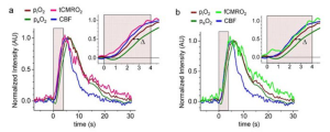
“We aim to develop a minimally invasive optical technique for real-time measurement of CMRO2 concurrently with cerebral blood flow (CBF),” write the investigators. “We used a pair of macromolecular phosphorescent probes with nonoverlapping optical spectra, which were localized in the intra- and extravascular compartments of the brain tissue, thus providing a readout of oxygen gradients between these two compartments. In parallel, we measured CBF using laser speckle contrast imaging.
“The method enables computation and tracking of CMRO2 during functional activation with high temporal resolution (~7 Hz). In contrast to other approaches, our assessment of CMRO2 does not require measurements of CBF or hemoglobin oxygen saturation.
“The independent records of intravascular and extravascular partial pressures of oxygen, CBF, and CMRO2 provide information about the physiological events that accompany neuronal activation, creating opportunities for dynamic quantification of brain metabolism.”

As noted above, the key step was to find a way to concurrently measure the difference between the extravascular and intravascular oxygen levels. For this, the researchers injected one phosphorescent probe, called Oxyphor PtR4, into the blood, and another probe, Oxyphor PtG4, was administered directly into the space between the blood vessels. The two Oxyphors have different colors, and by using two different lasers the team could measure the probes’ signals simultaneously with temporal resolution of ~7 Hz.
The technique was applied to a preclinical model, and the team demonstrated real-time computation and tracking of CMRO2 during functional activation of the brain. Using a third laser, the researchers also succeeded in measuring CBF in parallel with CMRO2 using the method called laser speckle contrast imaging.
“This clever development and use of probe pairs in the intra- and extravascular compartments opens the door to measurements of brain oxygen consumption,” said Andy Shih, PhD, a principal investigator at Seattle Children’s Research Institute’s Center for Developmental Biology and Regenerative Medicine


