
Human neural organoids, derived from pluripotent stem cells, enable the study of the highly regulated process of human brain development in vitro. In vivo, the process is shaped by strict time- and space-dependent morphogen gradients. Now, researchers have developed a method that leads to the development of a new cortex organoid that better represents the human brain.
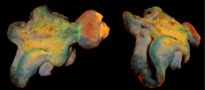
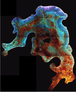
The new brain organoids are composed of a single, self organized apical-out neuroepithelium, ENOs (expanded neuroepithelium organoids), unlike standard brain organoid protocols in which multiple and independent neuroepithelium units (rosettes) are formed.
This work is published in Nature Communications, in the paper, “Temporal morphogen gradient-driven neural induction shapes single 3 expanded neuroepithelium brain organoids with enhanced cortical identity.”
The new findings show that the newly generated cortex organoid more closely represents the human brain in multiple aspects: their shape, their architectural organization, and several properties of their cells.
“The current brain organoid models generally have several developmental structures acting like an ‘independent’ small developing brain, within one organoid,” noted Benedetta Artegiani, PhD, research group leader at the Máxima Center. “Researchers have tried for a long time to mitigate the formation of these multiple structures. To diminish variability, increase reproducibility, better recreate the brain’s different cellular identities, and ultimately to recapitulate brain development more closely. Now in this research, the new organoid model is composed of this self-organizing, single developing neuroepithelium.”
[Benedetta Artegiani, Delilah Hendriks, Anna Pagliaro]
More specifically, the authors wrote that they found that “a prolonged, decreasing gradient of TGF-β signaling is a determining factor in ENO formation and allows for an extended phase of neuroepithelium expansion.”
Pediatric brain tumors may derive from an unusual or mistaken developmental process and their study could benefit from these new models that better recapitulate early embryonic developmental mechanisms. The signaling molecule that the researchers used to make the organoids is often altered in childhood brain tumors, also suggesting that the onset of cancer in young children could be linked with changes in brain development. Now that the researchers understand that temporal gradients are of great importance to generate more accurate organoids, this study paves the way to develop brain organoids more and more similar to the developing human brain.

