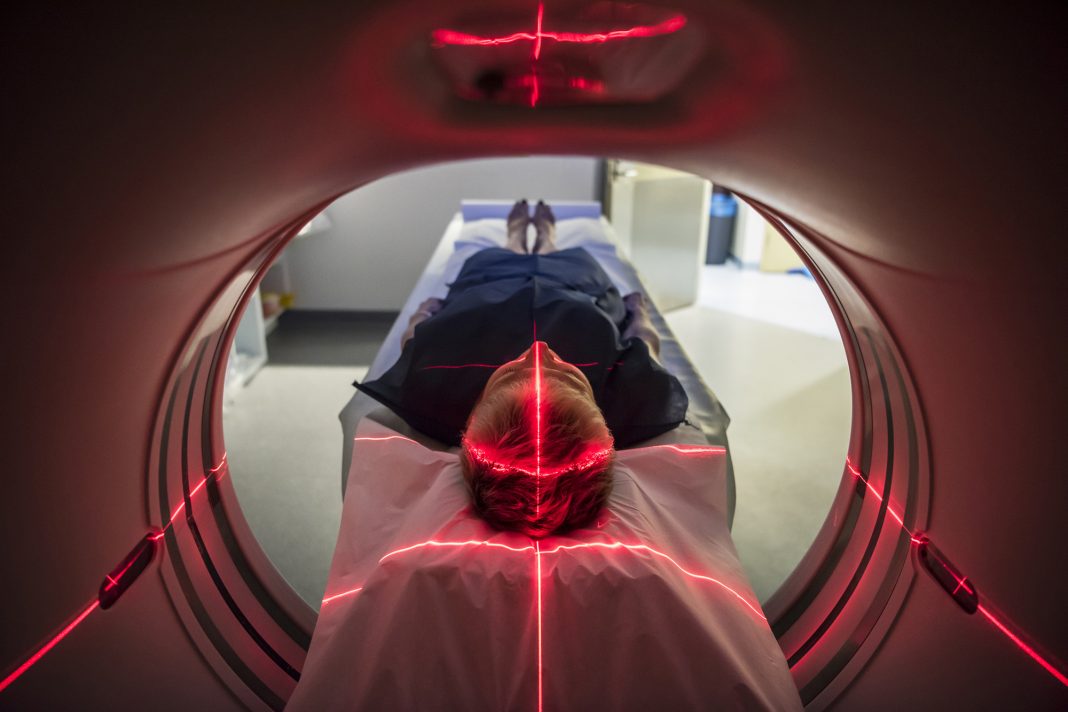
Discovery of a new radio-labeled molecule in a preclinical study that selectively reacts with high-energy radicals, a hallmark of focal innate immune activation, while turning a blind eye to radicals with lower redox potential that act as long-distance signals, could enable whole-body imaging through positron emission tomography or computed tomography (PET/CT) to detect multi-organ inflammation in real time, including manifestations of COVID-19 beyond the lungs and cytokine storm.
In a proof-of-concept study, the investigators used the new reporter molecule ([18F]4FN) in PET/CT imaging to discriminate among inflammatory foci in vivo in three mouse models of activated innate immunity: endotoxin-induced toxic shock, PMA-induced contact dermatitis, and lipopolysaccharide-induced ankle arthritis.
David Piwnica-Worms, MD, PhD, chair of Cancer Systems Imaging at the University of Texas MD Anderson Cancer Center, and the senior author of the study, has a long-standing interest in imaging myeloperoxidase (MPO) activity and innate immunity by exploiting the properties potential molecular scaffolds as magnetic resonance (MR) and PET imaging agents.

“Our approach focuses on interrogating the activation state of key participants of innate immunity, independent of specific cell type. [18F]4FN is an integrator of key cellular components of innate immunity and their individual cell activation states within this high redox oxygen radical-mediated inflammatory microenvironment,” said Piwnica-Worms. “Many alternative approaches monitor only cell trafficking or nonspecific low redox ROS [reactive oxygen species], which may not be sufficiently selective to report innate immunity activation states and production of high redox ROS.”
At present, there are no FDA-approved PET imaging agents capable of detecting high-energy ROS, resulting in an unmet clinical need to monitor the activation of innate immunity in vivo in different inflammatory diseases.
“Imaging highly reactive radical species by PET/CT is a step beyond imaging of cell trafficking which does not interrogate the activation states of the imaged cells,” said Piwnica-Worms. “Single cell surface markers can currently image a desired cell within a foci-of-interest, but the activation state of the cell within that site remains unknown. This decreases the diagnostic utility of the approach.”
The study was published in the journal Nature Biotechnology, in an article titled, “Imaging of innate immunity activation in vivo with a redox-tuned PET reporter.”
Innate immunity, driven by myeloid cells such as neutrophils, macrophages, and natural killer cells, is the body’s first line of defense against invading pathogens and acts non-specifically against foreign agents through pattern recognition receptors that are encoded in the germline. Myeloperoxidase (MPO), a highly conserved proinflammatory enzyme in the innate immune pathway across myeloid cells, is activated by hydrogen peroxide (H2O2) to produce a variety of high-energy radicals that work to eliminate pathogens.
“Innate immune cells reside constitutively in a resting state, but already primed with translated effector proteins lying in wait for stimulus-mediated activation. Standard sequencing and IHC data typically only illuminate cell localization, not activation,” said Piwnica-Worms.

The research team, including Federica Pisaneschi, PhD, assistant professor, and Seth Gammon, PhD, associate professor, both at Cancer Systems Imaging, used new techniques to synthesize [18F]4FN as a specific redox-tuned reporter of innate immune activity.
Piwnica-Worms said, “To facilitate broad adoption of [18F]4FN, a fully automated synthesis module provided a quick and efficient process compatible with the synthetic demands of short half-life 18F-labeled PET radiopharmaceuticals (~119 minutes). Using the relatively new copper-mediated deboronation/fluorination reaction of deactivated aryls, [18F]4FN was synthesized in good yields (>5% from end of bombardment), high radiochemical purity (>99%), and high molar activities (typically > 50 GBq/µmol, ‘tracer’ regime).” When oxidized by MPO and H2O2 , but not H2O2 alone, [18F]4FN binds selectively to nearby proteins and cells.

The team then evaluated the use of [18F]4FN as an in vivo PET imaging tool in several laboratory mouse models of inflammation. The molecule successfully highlighted innate immunity-mediated inflammation from acute toxic shock, arthritis, and contact dermatitis. Their results suggest [18F]4FN is a more specific and robust reporter of inflammation than other clinically utilized PET imaging agents, such as fluorodeoxyglucose ([18F]FDG).
“There is a lack of and an unmet need for imaging biomarkers to detect oxidative stress in patients. The demonstrated superiority of [18F]4FN over [18F]FDG in detecting inflammation shows great translational potential and applicability to detect oxidative stress and advance human health,” said John Chen, MD, PhD, a radiologist and investigator at Mass General Research Institute at Harvard Medical School, who is not part of the study.
Chen added, “While the broad sensitivity to a variety of oxidative species may limit its utility for testing and monitoring novel therapies against specific targets, it would be of great value in assessing the overall oxidative status of the patient and complement diagnostic methods with more specific targets.”
The oxidative burst that is characteristic of an activated innate immune response against microbial and viral infections, also contributes to other inflammatory diseases such as non-alcoholic steatohepatitis (NASH), cancer, atherosclerosis, and septic shock, increasing the range of potential applications for [18F]4FN.
“Future applications for [18F]4FN PET might include prognostic or pharmacodynamic imaging of these [inflammatory] diseases and therapy,” Piwnica-Worms said. “Clinicians currently manage these patients locally blinded to the state of multi-focal disease for which [18F]4FN PET may assist therapeutic decisions, including upstaging and triggers for aggressive treatment.”
“There is well documented histopathologic evidence for the infiltration of innate immunity at sites of immune-related adverse events, particularly in arthritis, hepatitis, colitis, thyroiditis, and hypophysitis, for which we hope to image the activity by [18F]4FN PET,” Piwnica-Worms added.
Once approved through clinical trials, physicians could use [18F]4FN PET/CT to identify hotspots of inflammation, enabling interventions before disease progression or monitoring patients in treatment.
Piwnica-Worms’ team is collaborating with clinicians to test specific applications of [18F]4FN. In an initial study, the team intends to evaluate [18F]4FN as an early biomarker of immune-related adverse events in patients being treated with immune checkpoint inhibitors. This proposed study is currently being reviewed by the FDA for Investigational New Drug (IND) registration and the approval of the Institutional Review Board (IRB).


