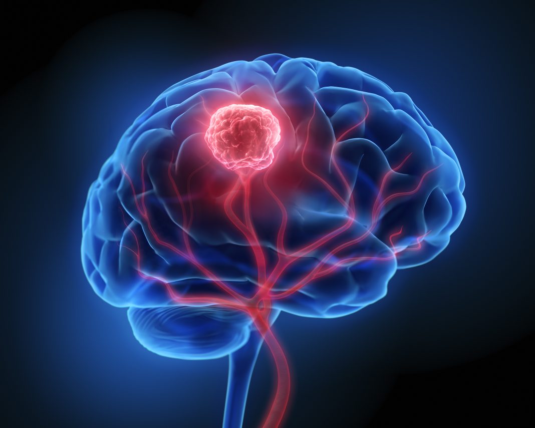
Researchers at the University of Bristol in the U.K. are exploring how mathematical modeling might be used in the development and use of blood-based biomarkers for brain tumors. The development of a simple blood test for glioblastomas (GBMs) could mean earlier diagnosis and more effective and personalized treatment options.
In a new study “Liquid biopsies for early diagnosis of brain tumors: in-silico mathematical biomarker modeling”, published in The Royal Society Interface, mathematical models were developed and paired with experimental data. The scientists found that for the prospective GBM biomarker Glial fibrillary acidic protein (GFAP) lowering the current biomarker threshold could lead to earlier detection of GBMs. The team also used computational modeling to explore the impact of tumor characteristics and patient differences on detection and strategies for improvements.
“Brain tumors are the biggest cancer killer in those under 40 and reduce life expectancy more than any other cancer. Blood-based liquid biopsies may aid early diagnosis, prediction, and prognosis for brain tumors. It remains unclear whether known blood-based biomarkers, such as glial fibrillary acidic protein (GFAP), have the required sensitivity and selectivity,” write the investigators. “
“We have developed a novel in silico model which can be used to assess and compare blood-based liquid biopsies. We focused on GFAP, a putative biomarker for astrocytic tumors and glioblastoma multi-formes (GBMs). In silico modeling was paired with experimental measurement of cell GFAP concentrations and used to predict the tumor volumes and identify key parameters which limit detection. The average GBM volumes of 449 patients at Leeds Teaching Hospitals NHS Trust were also measured and used as a benchmark.

This research is part of a wider University of Bristol-led CRUK project to develop an affordable, point of care blood test to diagnose brain tumors. This cross-disciplinary project combines biomarker discovery, development of fluorescent nanoparticle, and new testing techniques with computational modeling.
“Our findings provide the basis for further clinical data on the impact of lowering the current detection threshold for the known biomarker, GFAP, to allow earlier detection of GBMs using blood tests. With further experimental data, it may also be possible to quantify tumor and patient heterogeneities and incorporate errors into our models and predictions for blood levels for different tumors,” says Johanna Blee, PhD, lead author and research associate in the university’s department of engineering mathematics.
“We have also demonstrated how our models can be combined with other diagnostics such as scans to enhance clinical insight with a view to developing more personalized and effective treatments. These mathematical models could be used to examine and compare new biomarkers and tests for brain tumors as they emerge. We are hopeful this research will ultimately aid the development of a simple blood test for brain tumors, enabling earlier and more detailed diagnoses.”
To learn about other advances in brain cancer research see GEN: “Brain Cancer Therapy Shows Promise in Pediatric Clinical Trial” and “Researchers Uncover Dynamic Cells Connected to Brain Tumor Growth.”


