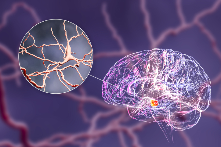
University of Arizona researchers have revealed new insights into the neurobiological cause of the involuntary, uncontrollable movement disorders that develop in patients with Parkinson’s disease (PD) after years of treatment with levodopa. Their study, in a rodent model, uncovered new findings about the nature of debilitating levodopa-induced dyskinesia (LID) that challenges current views. The also indicate how the anaesthetic ketamine could be used therapeutically to reduce LID by disrupting abnormal repetitive electrical patterns in the brain and allowing the motor cortex to regain control.
“By understanding the basic neurobiology underlying how ketamine helps these dyskinetic individuals, we might be able to better treat levodopa-induced dyskinesia in the future,” said Stephen Cowen, PhD, an associate professor in the department of psychology. Cowen is co-senior author of the team’s published paper in Brain, titled “Decoupling of motor cortex to movement in Parkinson’s dyskinesia rescued by sub-anaesthetic ketamine.” In their paper the team concluded, that their data “… further support the anti-dyskinetic properties of ketamine and suggest that ketamine acts to reduce LID by disrupting pathological interactions between motor cortex neurons during 10 dyskinesia.”
Parkinson’s disease is a neurological disorder of the brain that affects a person’s movement. The disorder develops when the level of dopamine, a chemical in the brain that’s responsible for bodily movements, begins to dwindle. “Parkinson’s disease (PD) is associated with debilitating motor and cognitive deficits resulting from the degeneration of midbrain dopaminergic neurons,” the authors explained. Administering the drug levodopa (L-DOPA), which is converted into dopamine in the brain, can help to counter disease-associated dopamine loss.
However, over a number of years, the brain of a Parkinson’s patient adapts to the levodopa treatment, and long term levodopa therapy can lead to dyskinesia, explained lead author Abhilasha Vishwanath, PhD, a postdoctoral research associate in the U of A department of psychology. “Uncontrolled movements are a defining and debilitating feature of LID,” the authors noted. So, while levodopa has been the gold-standard treatment for over 60 years, nearly 40% of PD patients develop levodopa-induced dyskinesia after 4-6 years of treatment. And while the excessive, uncontrolled movements associated with LID are debilitating, the team continued, “The neural generators of these movements are not understood, and treatments are limited.”
The chronic reduction in dopamine that occurs in PD alters what’s known as the cortico-basal ganglia-thalamic circuit in the brain, the team further explained. The brain’s motor cortex—the brain region responsible for controlling movement—is a key component of this circuit as it influences the activity of basal ganglia neurons. “Moreover, neuro adaptations of the basal ganglia in PD alter primary motor cortex (M1) excitability, synchrony, and the relationship between M1 activity and movement,” the researchers further stated. And M1 activity is also strongly affected by LID.
Through their newly reported study in a rat model of PD the researchers recorded activity from thousands of neurons in the motor cortex. “There are about 80 billion neurons in the brain, and they hardly shut up at any point,” Vishwanath commented. “So, there are a lot of interactions between these cells that are ongoing all the time”.
The study results indicated that these neurons’ firing patterns showed little correlation with the dyskinetic movements, suggesting that the motor cortex becomes essentially “disconnected” during dyskinetic episodes rather than direct causation. The findings, the investigators pointed out, “… suggest that primary motor cortex does not directly trigger specific dyskinetic movements during LID but, instead, dysregulated motor cortex activity may permit aberrant movements to spontaneously emerge in downstream circuits.” This finding challenges the prevailing view that the motor cortex actively generates these uncontrollable movements.
“It’s like an orchestra where the conductor goes on vacation,” said Cowen. “Without the motor cortex properly coordinating movement, downstream neural circuits are left to spontaneously generate these problematic movements on their own.”
The group noted that their previous preclinical work had shown that repeated administration of sub-anesthetic doses of ketamine could reduce established LID for weeks and attenuate the development of LID. “A clinical case study further supported anti-dyskinetic activity of ketamine and in a Phase I clinical trial (NCT06021756), both safety and tolerability were shown, and possible efficacy in individuals with LID was evident,” the authors added.
Through their newly reported study the researchers demonstrated that ketamine could help disrupt abnormal repetitive electrical patterns in the brain that occur during dyskinesia, which could potentially help the motor cortex to regain some control over movement. The collective results, the team noted, “… suggest that the relationship between M1 activity and movement changes considerably during LID, with M1 becoming effectively decoupled from sub-second changes in movement.” The findings, they added, indicated that “… aberrant movements in LID may not be directly initiated by M1 but indirectly initiated by way of reduced M1 inhibition of competing movements.”
Ketamine works like a one-two punch, Cowen said. It initially disrupts these abnormal electrical patterns occurring during dyskinesia. Then, hours or days later, ketamine triggers much slower processes that allow for changes in the connectivity and activity of brain cells over time—known as neuroplasticity—that last much longer than ketamine’s immediate effects. Neuroplasticity is what that enables neurons to form new connections and strengthen existing ones. And with one dose of ketamine, beneficial effects can be seen even after a few months, Vishwanath said.
The new study findings about motor cortex involvement in dyskinesia could point to new therapeutic approaches. They also gain additional significance in light of an ongoing Phase II clinical trial at the U of A, where researchers at the department of neurology are testing low doses of ketamine infusions as a treatment for dyskinesia in Parkinson’s disease patients. Early results from this trial appear promising, Vishwanath said, with some patients experiencing benefits that last for weeks after a single course of treatment.
Ketamine doses could be tweaked in a way such that the therapeutic benefits are maintained with minimized side effects, Cowen suggested.


