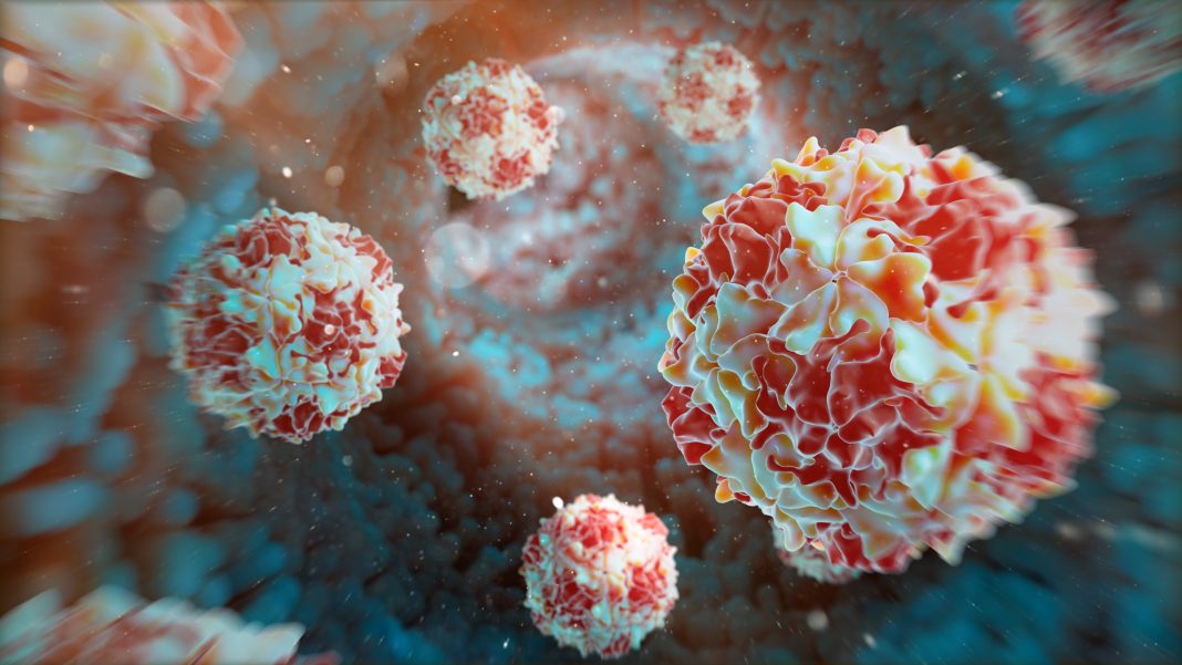
Scientists at Umeå University in Sweden report that they can now show how the poliovirus behaves when it takes over an infected cell and tricks the cell into producing new virus particles. Polio was thought to be almost eradicated, but infection has now been rediscovered in London and New York.
“We now have a completely different understanding of how the virus acts and thus better opportunities for research to perhaps find new ways to curb the virus’ progress in the future,” said Lars-Anders Carlson, PhD, in the university’s department of medical chemistry and biophysics, whose group, which included collaborators at the U.S. National Institutes of Health and Monash University in Australia, published its study “Membrane-assisted assembly and selective secretory autophagy of enteroviruses” in Nature Communications.
It has been known for some time that enteroviruses like the poliovirus drastically rearrange the inside of infected cells, but it has not been understood exactly how, because technology has not allowed us to see so deeply into the cells, according to the researchers who used a cryo-electron microscope to take three-dimensional images of how the poliovirus forms and takes over human cells.
“We were surprised to see how the virus transforms processes in the cell that are otherwise used to destroy viruses to produce new viruses instead,” added Carlson.
Identified the cellular site
The team was able to identify the site in the cell where the poliovirus forms new virus particles by seeing sites with half-assembled viruses. This “virus factory” in the cell turned out to be surfaces on the cell that resembled an otherwise normal process in the cell, autophagy, which serves to break down particles that the cell wants to remove, such as virus particles. But the poliovirus manages to reprogram this defense mechanism against viruses to produce more virus instead.
“Enteroviruses are non-enveloped positive-sense RNA viruses that cause diverse diseases in humans. Their rapid multiplication depends on remodeling of cytoplasmic membranes for viral genome replication. It is unknown how virions assemble around these newly synthesized genomes and how they are then loaded into autophagic membranes for release through secretory autophagy,” the investigators wrote.
“Here, we use cryo-electron tomography of infected cells to show that poliovirus assembles directly on replication membranes. Pharmacological untethering of capsids from membranes abrogates RNA encapsidation. Our data directly visualize a membrane-bound half-capsid as a prominent virion assembly intermediate. Assembly progression past this intermediate depends on the class III phosphatidylinositol 3-kinase VPS34, a key host-cell autophagy factor.

“These findings provide an integrated structural framework for multiple stages of the poliovirus life cycle.”
The researchers found that certain proteins are particularly important. The VSP34 protein is used by the virus to build new virus particles. When the researchers inhibited VSP34, they could see that the virus could barely assemble whole viruses, but mostly only half virus particles.
Another important protein is called ULK1, which slows down the production of viruses. The researchers could see that the amount of virus exploded when this protein was inhibited. This confirms the theory that the poliovirus breaks down this “brake.”
Once the virus has multiplied in the cell, the particles are released in vesicles to infect new cells. Here, the researchers also made a surprising discovery: a careful sorting of what is packed into the vesicles takes place. Only viruses that are correctly formed and carry the genetic material of the virus are placed in the vesicles, while empty virus particles are not allowed in. In this way, the virus may spread more efficiently.
“The new knowledge we are contributing about the role of autophagy in virus formation may provide new insights for the development of future antivirals that could complement vaccines,” noted Carlson. “We have good reason to believe that our findings are valid for the large group of viruses to which poliovirus belongs, enteroviruses. There is no vaccine against most enteroviruses, but an antiviral that acts on the autophagy system could be effective against many of them. However, there is still a long way to go.”


