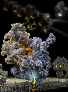
Researchers from Finland, Sweden, the U.K., and the U.S. say they have captured ribosomes translating messenger RNA (mRNA) expressed from the maternally inherited genome. The team which published its study “Mechanism of membrane-tethered mitochondrial protein synthesis” in Science, used cryo-electron microscopy group and discovered a novel mechanism that mitochondrial ribosomes use for the synthesis and delivery of newly made proteins to prevent premature misfolding.
Disruptions of this process underlie a large group of human diseases that can strike at any age, differing in the affected tissue and severity. However, the molecular mechanisms of these processes have remained obscure. But the advance in biological imaging brought about by cryo-electron microscopy now provides researchers with the tools to investigate the functions of individual proteins at unprecedented resolution and detail, according to the scientists.
“Mitochondrial ribosomes (mitoribosomes) are tethered to the mitochondrial inner membrane to facilitate the cotranslational membrane insertion of the synthesized proteins. We report cryo–electron microscopy structures of human mitoribosomes with nascent polypeptide, bound to the insertase oxidase assembly 1–like (OXA1L) through three distinct contact sites. OXA1L binding is correlated with a series of conformational changes in the mitoribosomal large subunit that catalyze the delivery of newly synthesized polypeptides,” write the investigators.
“The mechanism relies on the folding of mL45 inside the exit tunnel, forming two specific constriction sites that would limit helix formation of the nascent chain. A gap is formed between the exit and the membrane, making the newly synthesized proteins accessible. Our data elucidate the basis by which mitoribosomes interact with the OXA1L insertase to couple protein synthesis and membrane delivery.”
The capture of the mitochondrial ribosome translating mRNA into a protein revealed a gating mechanism to prevent newly made proteins from prematurely misfolding. For proteins to be functional within our cells requires coordinated folding processes to obtain a correct 3D shape. Disruptions to protein folding can have profound biological implications for all organisms and in humans lead to devastating diseases.
“Getting a direct picture of a biological process that we investigated for several years by biochemical and genetic tools is absolutely electrifying,” says Uwe Richter, PhD, a shared first author and PI at the University of Helsinki and Newcastle University.
“This study highlights the power and brilliance of international collaborative basic science driven from the bottom-up,” adds Brendan Battersby, PhD, research director, Institute of Biotechnology, University of Helsinki and one of the corresponding authors. “There is a worrying trend among scientific funding agencies to direct research from the top towards goal-oriented tasks pursued by large consortia, and in the process deprive research funds away from individual investigators who are the cornerstone of scientific discovery. The best scientists will always seek each other out to follow the creativity of their ideas, which the track record shows unquestionably leads to real innovation in the end.
“Understanding the fine details of these cellular mechanisms has important considerations for human diseases but also for the side-effects of commonly prescribed antibiotics. [The gene expression of mitochondria] has many overlaps with that of bacteria and as a result many antibiotics used to treat bacterial infections can also disrupt our cellular powerhouses, accounting for side-effects of these medications.”
“Solving these ribosome structures is integral to the development of effective and safe new antibiotics in the future. In the end, this highlights the importance of basic bottom-up research and how it continues to drive innovation and we cannot afford to lose it.”


