Technologies for each of the “omes”—the genome, proteome, transcriptome, epigenome, metabolome, and so on—are in the middle of their own individual revolutions. As these technologies mature, they not only facilitate the exploration of their respective omes, but they also create opportunities to relate one ome to another. Indeed, rather than use standalone omics technologies, researchers may prefer to use multiomics technologies. With the latter, researchers may combine different kinds of omics data to conduct more comprehensive studies of cells and tissues.

BioSkryb Genomics
Traditionally, multiomics has layered one ome on top of another. That is, multiomics has relied on computational tools to correlate information from one omics dataset to another omics dataset. Many of the technologies that people refer to as multiomics technologies infer “what is happening in the genome because they are looking at a small area, a minute end of a transcriptome,” says Reagan Tully, chief commercial officer, BioSkryb Genomics.
But some technologies aim to measure different omes simultaneously. “While there are many approaches and technologies of value for biological measurement,” notes Anjali Pradhan, senior vice president, product management and marketing, Mission Bio, “simultaneous multiomics measurement enables clinically actionable insights that cannot be derived by bulk next-generation sequencing or RNA sequencing approaches alone.”
For example, as Pradhan adds, measuring cell genotype and immunophenotype together, via proteogenomic profiling, can unlock new understandings of tumor heterogeneity. And adding the ability to interrogate samples at single-cell resolution expands the reach of multiomics even further, opening applications from the research laboratory to the clinic.
Their favorite ome is the proteome
Last May, Inside Precision Medicine (GEN’s sister publication) held an online event called “The State of Precision Medicine.” One session focused on multiomics, specifically, the importance of adding proteomics data to genomics information. Prominent speakers included Jennifer Van Eyk, PhD, professor of medicine at Cedars-Sinai Medical Center, and Mathias Uhlén, PhD, professor of microbiology at KTH School of Engineering Sciences in Chemistry, Biotechnology, and Health. They referred to themselves as “protein fanatics.”
A patient’s genome, Van Eyk noted, contains information about that patient’s disease predispositions and drug responses. She added, however, that better information about disease risks and drug responses could be gleaned from the proteome. Although there are only so many protein-encoding genes, the intricacies of protein expression generate various kinds of proteomic information in abundance. According to Van Eyk, information about disease-induced modifications, isoforms, concentration changes, and chemical complexity can inform predictions of what will happen in the body, in the context of the body and the environment. She suggests that a proteomics approach—one that would involve monitoring of not just one protein at a time, but thousands—could generate valuable clinical insights.
Uhlén emphasized the importance of proteins as drug targets. After noting that the field of antibody-based proteomics had enjoyed explosive growth over the last two to three years, he declared that the growth would go on, and that more data would be produced more quickly. And after emphasizing that proteomics technology can identify drug targets independently or in combination with other omics technologies, Uhlén gave one more prediction: “We are entering the most interesting era in medical research ever.”
Creating complete omes
Jay West, PhD, chief technology officer at BioSkryb Genomics, likely agrees with Uhlén’s last prediction. In fact, he may be inclined to take the prediction a step further by channeling Peter Drucker, the management consultant who famously said, “The best way to predict the future is to create it.”

BioSkryb Genomics
West has had a standing interest in single cells. When West was working on his doctorate in molecular pharmacology at the University of California, Davis, he isolated single cells using microscopes and analytical chemistry. Then he moved into industry, taking a position at Fluidigm (now Standard Biotools), where he was part of building a single-cell technology platform.
During that time, West met Chuck Gawad, MD, PhD, then a postdoc in the laboratory of Stephen Quake, DPhil, professor of bioengineering and applied physics at Stanford University. Quake is also a co-founder of Fluidigm. Gawad and West stayed in touch, even when Gawad moved to start his own laboratory at St. Jude Children’s Research Hospital, where he invented primary template-directed amplification (PTA) technology. PTA is a whole-genome amplification technology that allows for robust sequencing coverage and variant detection even in small samples such as single cells.
In addition to sharing an interest in single cells and genomics technologies, both Gawad and West were highly motivated to work in cancer and to try to make a difference. Gawad became a pediatric oncologist and is currently a Chan Zuckerberg Biohub Investigator, as well as an associate professor at Stanford University. Together, in 2018, Gawad and West co-founded BioSkryb Genomics and licensed the PTA technology from St. Jude.
 Before long, they were sequencing clinical patients’ samples. One of the lessons they learned early on, West tells GEN, is that a lot of data is missing that could be impactful for the patient. “If you are reading two pages out of a 500-page book,” he says, “it’s hard to get the whole story.”
Next-generation sequencing is great for detecting germline differences, but it is not great at detecting somatic variation between cells, West notes. And that is where disease is found. So, the two decided to focus on comprehensive single-cell omes.
“Omes, to us, means all, not partial” West stresses. “We strive to create complete omes.” They started with genomes. And now, building off the genome amplification chemistry, they have been working to layer on additional omes.
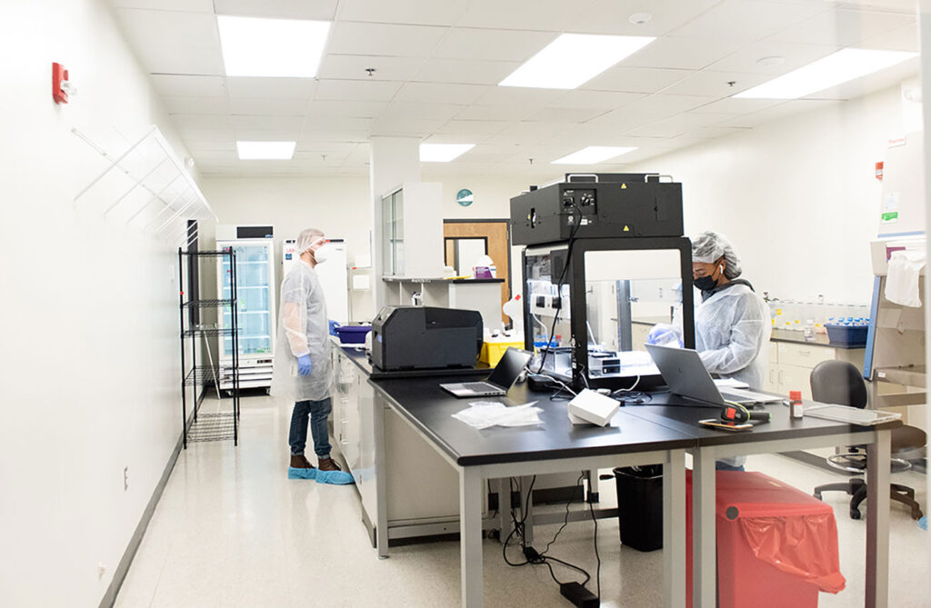
When COVID-19 hit, most of Bioskryb Genomics went home to socially isolate. But a very small group stayed in the laboratory to figure out how to build a product that layered the transcriptome on top of the genome. This work bore fruit earlier this year, when the company launched ResolveOME, which combines transcriptome and genome information. The ResolveOME workflow includes the following steps: isolation of single cells; cytosolic lysis and reverse transcription; nuclear lysis and genomic amplification (via the PTA technology); fraction separation and the construction of RNA and DNA libraries; and multiomics analysis.
Now that Bioskryb Genomics has combined the genome and transcriptome, the company is moving into the proteome. At the American Association for Cancer Research meeting earlier this year, the company released protein analysis data, with transcriptome and genome information, from 15 clinical patient samples. West says that the company is heading toward the consolidation of four omes within the next 18 months, having set its sights on the epigenome.
How many omes is enough? West says that’s a good question, but he keeps his focus on whatever is needed to help patients.
Omes in space
The multiomics wave and the spatial biology wave are overlapping. The result: an unusually impressive example of constructive interference. The RNA-based spatial platforms are working on detecting proteins, and the protein-based platforms are adding transcriptomes to their offerings.
“There is an obviousness on why we need to do multiomics analysis on most of our samples, but I believe the obviousness is most true in spatial analysis,” notes Niro Ramachandran, PhD, chief business officer, Akoya Biosciences.

A major requirement in spatial analysis, he adds, is the need to identify cell boundaries, a need that does not exist in nonspatial analysis. Spatial proteomics analysis is best suited to identify cell boundaries and cell type. Spatial transcriptomics approaches allow exhaustive analysis of cell function. The combination provides the most powerful example of multiomics analysis.
One stumbling block is being able to do both analyses simultaneously on the same tissue slide. In many cases, staining for two omics means using two serial sections.
To enable the detection of RNA and protein in the same tissue, Akoya Biosciences partnered with Advanced Cell Diagnostics (a Bio-Techne brand) to develop a single-cell, spatial multiomics workflow. This workflow, which was introduced last year, encompasses Akoya Biosciences’ PhenoCycler-Fusion protein imaging assays and an automated version of Advanced Cell Diagnostics’ RNAScope HiPlex assay for RNA imaging. The RNAScope assay can allow single-molecule detection of up to 12 RNA targets simultaneously.
Also, in the past year, Vizgen unveiled a protein co-detection kit, enabling subcellular spatial multiomics measurement with the simultaneous detection of RNA and proteins during a standard multiplexed error-robust fluorescence in situ hybridization (MERFISH) experiment.
But is it too early to combine these two technologies, given that they are relatively immature? Jasmine Plummer, PhD, director of spatial omics at St. Jude Children’s Research Hospital, says that the technologies are “new and exciting but need more time to be vetted.” She adds that it makes sense to “focus on one species, be it RNA or protein, until we fully understand how these technologies are performing.”

Spatial biology companies usually focus on the transcriptome or the proteome. Bioskryb Genomics, however, intends to take a different approach. Although Bioskrb is moving into spatial biology, the company wants to do so while focusing on the genome.
Bioskryb uses what West calls a “really fancy colony picker” (through a partnership with Sartorius) to pick cells out of a tissue section. From there, the company can run multiomics analyses on the cells.
West says that when you think about invasive cancer, you want to know why the cancer is invading. You should be aware, he continues, that “looking at 40 markers of the transcriptome is not going to tell you that.” He says that because cancer is a genetic disease, you “need to know what is going on in the genome.”
When you’re doing deep genomics in cancer, West adds, you’re not just looking at a spatial image, you’re looking at a temporal change. The normal cells are dominating the tissue section. But there are later-stage cells with genomic instability that instigate invasive cancer. Referring to some unpublished data, West tells GEN that Bioskryb has been able to track the progression of cells—an “incredible” feat, he says, and one of the most rewarding things the company has done so far.
But the utility of multiomics goes far beyond cancer. Another critical area, Pradhan notes, is characterization of gene and cell therapies, where both the safety and efficacy can depend on efficiently characterizing transduction efficiency, vector copy number, and integration sites.
And though it’s still early days for multiomics—and challenges like large amounts of data, cost, accessibility, and more, will all need to be ironed out—the excitement lies in the technology’s ability to uncover new biological understandings that would otherwise remain out of reach.


