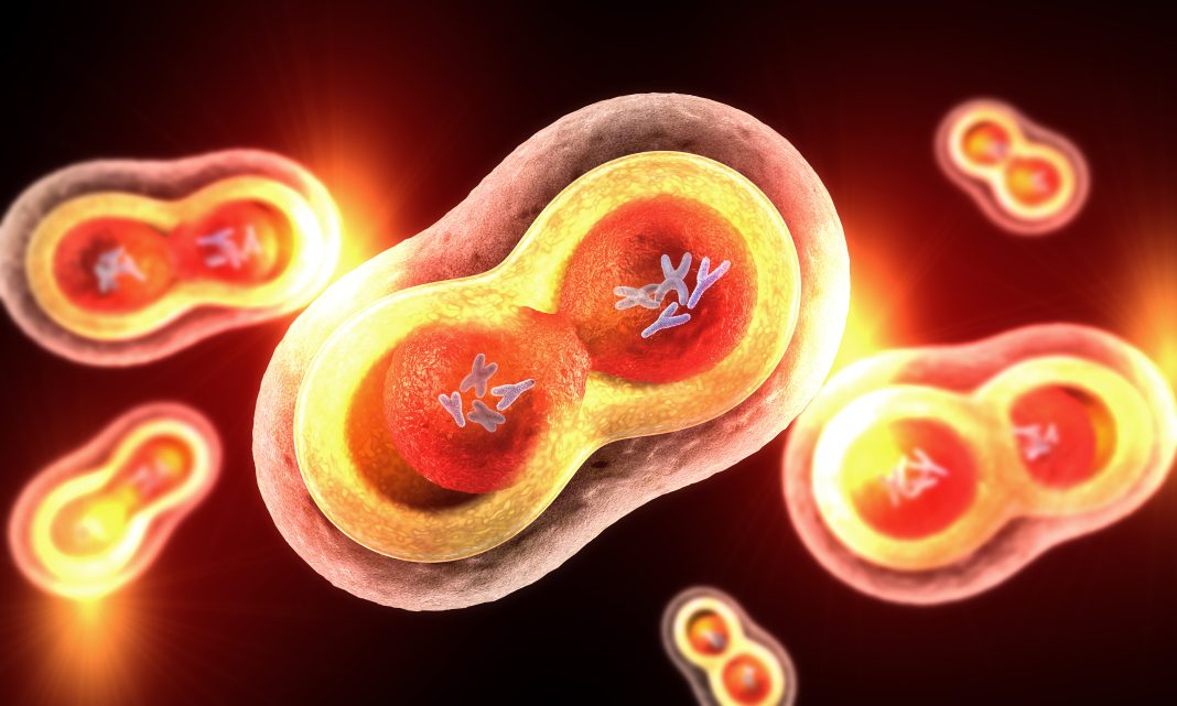
New findings shed light on how chromosomes manage the various pushing and pulling forces generated when cells divide. Specifically, researchers show that chromosomes resist being punctured by the microtubules that are responsible for the pushing by a chemical modification known as deacetylation. This modification helps ensure that exactly one chromosome copy is distributed to each of the two daughter cells.
The work was led by Daniel Gerlich, PhD, a senior scientist at the Institute of Molecular Biotechnology of the Austrian Academy of Sciences. His team’s findings were published in the journal Nature (“A mitotic chromatin phase transition prevents perforation by microtubules”).
When cells divide by mitosis, they condense long DNA molecules into structures called chromosomes so that they can be transported by the spindle apparatus, which is composed of thousands of microtubules. Certain microtubules cause what are known as polar ejection forces that push chromosome arms toward the middle of the dividing cell—and away from the spindle poles. The chromosome arms must resist the polar ejection forces and prevent the microtubules from penetrating them.
Gerlich’s team showed that an absence of acetyl groups on histones, or structural proteins in the chromosomes, was the key to this resistance. “Here we show how global histone deacetylation at the onset of mitosis induces a chromatin-intrinsic phase transition that endows chromosomes with the physical characteristics necessary for their precise movement during cell division,” they wrote in their article.
The researchers treated cells with the drug trichostatin A (TSA), which inhibits enzymes that remove acetyl groups from the histones. TSA treatment led to hyperacetylation of the histone tails and decompaction of the chromosomes. This caused the microtubules to puncture the chromosome surface.
“Deacetylation-mediated compaction of chromatin forms a structure dense in negative charge and allows mitotic chromosomes to resist perforation by microtubules as they are pushed to the metaphase plate. By contrast, hyperacetylated mitotic chromosomes lack a defined surface boundary, are frequently perforated by microtubules and are prone to missegregation,” summarized the researchers.
Condensins have long been known to play a role in chromosome structure, but the details have been unclear. When the researchers treated condensin-depleted cells with TSA, the chromatin was distributed throughout the cytoplasm. This indicates that, in the absence of condensin, histone deacetylation causes complete compaction of mitotic chromosomes.
“The current study could have implications for our understanding of how condensin is involved in chromosome packaging,” wrote Kazuhiro Maeshima, PhD, a professor in the Structural Biology Center at the National Institute of Genetics in Japan, in a piece that accompanied the research article. “Although the team shows that histone deacetylation can induce complete compaction of mitotic chromosomes in the absence of condensin, the shape of the chromosomes is aberrant. To make a proper rod-like chromosome shape, then, loop formation by condensin seems crucial.”
“Future work combining sophisticated imaging techniques, computational modeling and a technique called Hi-C that captures information about chromosome conformation could shed light on the mechanism of loop formation required to make the rod-like shape of mitotic chromosomes,” he added.
“Our study shows how DNA looping by the condensin complex cooperates with a chromatin phase separation process to build mitotic chromosomes that resist both pulling and pushing forces exerted by the spindle. The deacetylation of histones during cell division hence confers unique physical properties to chromosomes that are required for their faithful segregation,” concluded Gerlich.


