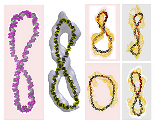
![DNA structures calculated with a supercomputer (in color) superimposed upon those derived from cryo-electron tomography data (in white or yellow). Each structure contains around 300 base pairs, but a different degree of winding. [Thana Sutthibutpong]](https://genengnews.com/wp-content/uploads/2018/08/Oct13_2015_ThanaSutthibutpong_SupercoiledDNA5918422010-1.jpg)
DNA structures calculated with a supercomputer (in color) superimposed upon those derived from cryo-electron tomography data (in white or yellow). Each structure contains around 300 base pairs, but a different degree of winding. [Thana Sutthibutpong]
Biologically active DNA is so dynamic and executes such extreme twists and bends that the nucleus is more Cirque du Soleil than parade ground. This statement may seem to be at odds with DNA’s iconic image, the upright double helix, but when it is tightly packed within the nucleus, DNA is supercoiled, not “at attention.”
Supercoiled DNA’s act was captured through the lens of cryo-electron tomography by researchers at the Baylor College of Medicine. These researchers, led by Lynn Zechiedrich, Ph.D., saw that when supercoiled DNA is wound and unwound, turn by turn, the molecule can assume a startling variety of poses, including previously unobserved splits—the separation of base pairs.
The Baylor researchers examined DNA minicircles, each of which contained about 300 base pairs. Each minicircle had a known degree of coiling. Moreover, each minicircle was deemed to be biologically active because it was shown to respond to human topoisomerase II alpha, an enzyme that manipulates DNA.
“Previous studies were on short fragments (6 to 12 base pairs) of linear DNA, but human DNA is constantly moving around in your body—and it coils and uncoils,” said Dr. Zechiedrich. “You can't coil linear DNA and study it, so we had to make circles so the ends would trap the different degrees of winding.”
The Baylor team’s cryo-electron tomography work was confirmed by computer simulations, which were conducted at the University of Leeds. Together, the Baylor and Leeds researchers published their work October 12 in Nature Communications, in an article entitled, “Structural diversity of supercoiled DNA.”
“Our results reveal that each topoisomer, negative or positive, adopts a unique and surprisingly wide distribution of three-dimensional conformations,” the authors wrote. “Moreover, we uncover striking differences in how the topoisomers handle torsional stress. As negative supercoiling increases, bases are increasingly exposed. Beyond a sharp supercoiling threshold, we also detect exposed bases in positively supercoiled DNA.”
The authors took note of how DNA could take many different shapes. According to the study’s co-lead author, Baylor’s Rossitza N. Irobalieva, Ph.D., “Some of the circles had sharp bends, some were figure-eight’s, and others looked like racquets or sewing needles. Some looked like rods because they were so coiled.”
“Being able to observe individual DNA circles allows us to understand the different structures of biologically active DNA. Each of these different structures facilitates how DNA interacts with proteins, other DNA and RNA, and anticancer drugs, adapting to the cell processes required,” said Jonathan Fogg, Ph.D., the other lead author of the publication, also of Baylor College of Medicine. “The next step is to start adding the other components of the cell or anticancer drugs to see how the DNA shapes change.”
Not only does the study provide clues for drug development, given that drugs must recognize particular DNA conformations to interact with DNA, it also helps researchers understand how a meter-long strand of DNA manages to fit within a cell’s nucleus. While the DNA minicircles used in the study are fairly small, about one ten millionth the size of a complete DNA strand, they appear to undergo splitting of a sort that can allow flexible hinge movements, accounting for the sharp bending that must occur in tightly packed DNA. Contortionists who cram themselves into knee-high boxes call this sort of thing enterology. But it seems that DNA is the ultimate enterologist.


