Sponsored content brought to you by
Advances in cell biology, neurology, oncology, and other disease-based research require an understanding of the spatial dynamics and interplay between heterogeneous cellular populations. Understanding the molecular basis of disease pathology or tissue homeostasis relies on knowing how thousands of biomolecules, including proteins, nucleic acids, lipids, and metabolites, interact in cells and tissues.
However, existing tools that offer multiomics capabilities are unable to fully unleash the power of spatial omics, or provide a comprehensive, integrated biological picture of the diverse array of molecules, including metabolites and lipids, that interact across long and short distances within cellular systems. These tools also cannot fully visualize the lipidome, metabolome, and glycans, all of which would offer a broader molecular insight and stronger basis for classification utilizing computational tools. Other issues include limited fields-of-view within a reasonable analysis time and intermediate plex levels that cannot track a sufficient number of proteins.
Only mass spectrometry (MS) can bring together a true multiomic experiment that informs on what makes changes happen biologically (via the proteome) and what has happened and is happening (via the metabolome and phenome).
MS requires only a single tissue section to map hundreds of biomolecules such as metabolites, lipids, and drug compounds in a label-free, untargeted manner. Yet, current MS-based protein expression imaging requires shotgun techniques where all proteins in a tissue are proteolyzed and individual proteolytic fragments are analyzed and lose the spatial dimension. This effectively limits speed, resolution, and the ability to rapidly multiplex targeted proteins of interest as well as reproducibility. While bulk proteomics is a valuable and necessary tool, having additional ways to visualize the spatial dimension is important.
A new approach for multiplexed protein profiling
Matrix-assisted laser desorption/ionization (MALDI) Imaging is widely used to obtain highly multiplexed images of nontargeted endogenous biomolecules from tissues. A new method, termed MALDI HiPLEX-IHC, enables highly multiplexed immunohistochemistry (IHC) to image targeted large intact biomolecules such as proteins in a straightforward, multiomic workflow.
IHC is broadly used to determine the structural organization of proteins by utilizing the specificity of antibodies, but can usually visualize only five or six tags at a time through fluorescence microscopy due to the intrinsic nature of molecular fluorophores that typically exhibit relatively broad excitation and emission bands.
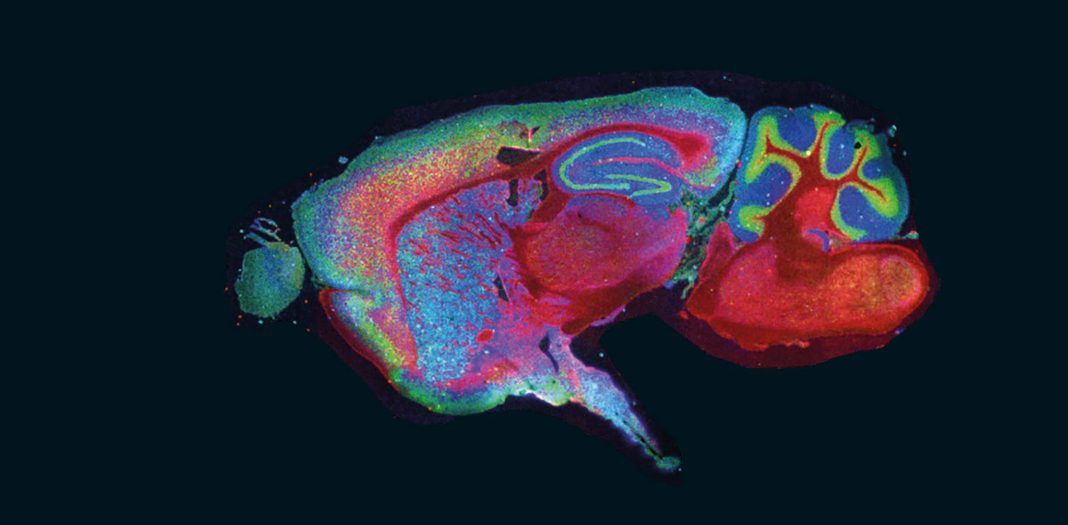
The MALDI HiPLEX-IHC workflow combines MALDI Imaging with IHC and can analyze, without cycling, over a hundred biomarkers in a single scan since each probe has a unique mass. This generates highly multiplexed images that can be combined with other imaging techniques for multimodal data, or subsequent MALDI Imaging runs for multiomic data visualizing small molecules, lipids, glycans, intact proteins, and other entities from the same tissue section.
Scientists can now co-localize small-molecule drugs and drug targets, such as their receptors, as well as associated biomolecules involved in the cellular response to a drug. Metabolic pathways associated with known proteins of interest can be spatially mapped and protein activity can be assessed quantitatively.
“HiPLEX-IHC combines the gold standards of tissue characterization—fluorescence microcopy and histopathological staining—with chemical-information-rich MALDI Imaging of lipids, small molecules, and proteins associating the large tissue imaging databases with unmatched discovery science,” said Jonathan V. Sweedler, PhD, professor of Chemistry, the University of Illinois Urbana-Champaign. “It is exciting to consider the new knowledge that awaits.”
Other advanced fluorescence techniques developed to achieve higher multiplexing are complex and laborious, and incomplete cycling can confound the results. In addition, methods such as multiplexed ion beam imaging (MIBI) and imaging mass cytometry (IMC) are limited by slow scan speeds and require specialized instrumentation unavailable in most labs.
Spatial proteomics utilizing MALDI Imaging has a wider field-of view for faster and less expensive analysis. Comparatively, MALDI Imaging is also less destructive and allows for multiple analyses on the same tissue section for fast data acquisition. An entire microscope slide can be profiled in under an hour at a spatial resolution of 40 µm.
Specialized dual-labeled probes with fluorophores
MALDI HiPLEX-IHC uses novel photocleavable mass-tag probes (PC-MTs), known as Miralys™ probes, available from AmberGen. The PC-MT works by staining tissues using the probes in a standard IHC workflow. A binding agent anchors the probe to the target molecule, and after staining, ultraviolet light cleaves the built-in PC-Linker. After application of the MALDI matrix, the specimen is placed in the MS, which detects the short peptide mass-tag attached to the PC-Linker, revealing proteins of interest at each location in the tissue section. Capture agents can include antibodies, lectins, and oligonucleotides.
A dual-labeled Miralys probe with a fluorophore attached to the binding agent emits both a fluorescence signal and a mass-tag signal. The PC-MT and the fluorophore show the position of the same molecule, enabling straightforward spatial co-registration of images. This is particularly valuable for researchers currently using fluorescence imaging. In one simple step, they can add molecular information without compromising the number of biomarkers that they can visualize.
Crucially, this highly multiplexed technology allows measurement of small molecules (such as metabolites), intact proteins, and other macromolecules in one workflow on the same formalin-fixed paraffin-embedded (FFPE) or fresh-frozen (FF) thin-tissue section.
“All-in-one systems are important for multiomics approaches, which are the future of tissue interrogation and biological science,” emphasized Elizabeth K. Neumann, PhD, assistant professor of Chemistry, UC Davis. “The ability to lean on well-established antibodies without having to cycle or bleach is awesome and will enable more difficult/brittle systems to be analyzed.”
The versatility of the approach opens opportunities to perform both label-free untargeted small molecule MALDI Imaging and multiplex PC-MT-based targeted MALDI Imaging of macromolecular biomarkers on the same tissue section, over >1 cm2 regions, with the option to subsequently concentrate on a smaller area of interest at higher resolution.
Opening the door to new knowledge
MALDI Imaging is increasingly relied upon to characterize the tumor microenvironment’s heterogeneity by determining distinct tumor subpopulations and understanding localized molecular changes in detail. In-depth spatial multiomic insights with MALDI HiPLEX-IHC can help researchers identify biomarkers for cancers and classify heterogeneous tumor subpopulations, to gain important contextual clues to tissue-level communication networks that are integral to cancer growth and treatment success.
“From the perspective of a lab heavily invested in cellular signaling processes involved with cancer, MALDI HiPLEX-IHC is a game changer allowing integration of mass spectrometry imaging with cell biology,” said Peggi Angel, PhD, associate professor of Cell & Molecular Pharmacology & Experimental Therapeutics, Medical University of South Carolina. “We will be using this technology for multiomic N-glycan and collagen imaging studies to understand aggressive breast cancers.”
MALDI HiPLEX-IHC, which is ideally suited to neurological and pathology research as well as archival tissue analysis, fits seamlessly into existing MALDI workflows for Bruker’s timsTOF fleX and rapifleX® platforms. Full software automation through SCiLS™ autopilot allows for “pushbutton” workflow setup, ensuring reproducible and consistent preparation without the need for highly trained experts. The targeted PC-MT probe technology results in clean MALDI spectra for easy readout.
Summary
By incorporating the advantages of MALDI Imaging, such as the label-free spatial mapping of many different metabolites, together with time-tested and trusted methods like IHC, MALDI HiPLEX-IHC allows researchers to perform highly multiplexed protein expression imaging with well-established MALDI instrumentation and workflows. This new spatial proteomic tool is set to transform spatial biology and the understanding of tissue homeostasis and disease pathology.
Transform your spatial omics with MALDI HiPLEX-IHC Imaging
www.bruker.com/maldi-hiplex-ihc.html


