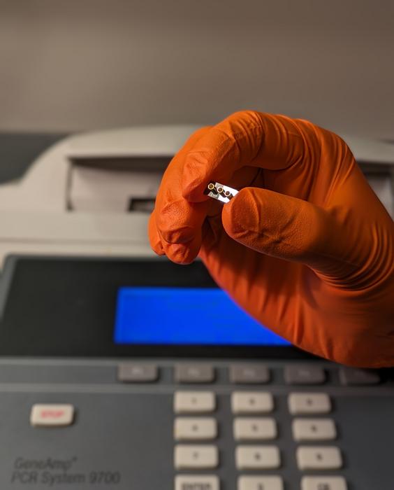
Researchers headed by a team at the University of California (UC), Santa Cruz, and at Brigham Young University, reported on their development of a lab-on-a-chip diagnostic system that combines optofluidics with nanopore technology to enable label- and amplification-free quantification of viral RNA from biofluids such as urine, whole blood, and throat swabs. The team showed that the platform could test for SARS-CoV-2 and Zika virus within hours using samples from primates, and with accuracy equivalent to, or even higher than high-precision PCR.
The researchers hope the technology will represent a major innovation for the future of rapid diagnostics. “This could turn into the next big diagnostic system,” said Aaron Hawkins, PhD, a professor of electrical and computer engineering at Brigham Young University. “You get sick, you go to the hospital or doctor, and their tests rely on this technology. There’s a path where this could be installed right there [in a hospital or clinic], so you wouldn’t have to wait to get your results.”
The new technology is the result of a longstanding collaboration between Hawkins, Holger Schmidt, PhD, UC Santa Cruz distinguished professor of electrical and computer engineering, and Jean Patterson, PhD, at the Texas Biomedical Research Institute. The team’s published report in PNAS is titled, “Label-free and amplification-free viral RNA quantification from primate biofluids using a trapping-assisted optofluidic nanopore platform.” In their paper, the team concluded, “The versatility, performance, simplicity, and potential for full microfluidic integration of the amplification-free nanopore assay points toward a unique approach to molecular diagnostics for nucleic acids, proteins, and other targets … These devices can find applications as POC diagnostics, research tools for assisting in the development of animal models, and many other fields.”
Over recent decades there have been multiple viral pandemics, including swine flu, Ebola, Zika, and COVID-19, creating the need for fast, accurate diagnostics, the authors wrote. “As preventive measures such as vaccinations and antiviral medicine are often either limited or unavailable, it is essential to develop simple, low-cost, and low-complexity point-of-care (POC) diagnostic technologies with high sensitivity, speed, and accuracy.”
However, while existing and inexpensive antigen tests such as enzyme-linked immunosorbent assays (ELISAs) are suitable for POC use, the team continued, “… they suffer from poor sensitivities and reliabilities when the biomarker concentration is low.” As a result, PCR testing remains the gold standard of accuracy for virology testing. But the method falls short in several ways. “PCR is complex and requires expensive reagents, central laboratory infrastructure, and well-trained personnel, making it ill-suited for use in low-resource environments,” the investigators stated. It can also sometimes take days to get testing results back.
These complex PCR reactions are needed for the amplification of viral DNA or RNA, a process of making multiple copies of the genetic material that can introduce and amplify error. PCR tests can also only detect nucleic acids, whereas for some diseases, it might be useful to be able to detect other biomarkers such as proteins.
Schmidt, Hawkins, and Patterson and their teams have now developed a diagnostic tool that solves PCR-related drawbacks. The new technology requires little sample preparation and is amplification free and label free—so does not use light to identify biomarkers. These features dramatically cut down the time and complexity of the diagnosis process, the team believes. “The potential is enormous,” Patterson said. “The idea that you don’t have to amplify to get accurate results is a huge advance, on par with how PCR was an incredible step forward when it came out.”
The new diagnostic system combines Schmidt’s area of expertise in optofluidics, which is the control of tiny amounts of fluids with beams of light, with a nanopore for counting single nucleic acids to read genetic material. The tool was designed to test for Zika and SARS-CoV-2, which have been priority areas for National Institutes of Health, which funded the research.
“We built up a simple lab-on-a-chip system that can perform testing at a miniature level with the help of microfluidics, silicon chips, and nanopore detection technologies,” said Mohammad Julker Neyen Sampad, Schmidt’s graduate student and the paper’s first author. “Simple, easy, low resource tool development was our goal—and I believe we got there.”
To run the test, a sample of biofluid is mixed in a container with magnetic microbeads. For this study, the researchers used biofluids including saliva and blood from baboons (SARS-CoV-2) and marmosets (for Zika virus) at Texas Biomedical Research Institute.
The microbeads are designed with a matching RNA sequence of the disease for which the test is designed to detect. So, for a COVID-19 detection test, the microbeads will have strands of SARS-CoV-2 RNA on them. If there is SARS-CoV-2 virus present in the sample, the virus’s RNA will bind to the beads. After a brief waiting period, the researcher pulls the magnetic beads down to the bottom of the container and washes everything else out.
The beads are put into a silicon microfluidics chip designed and fabricated by Hawkins’ group, where they flow through a long, thin channel covered by an ultra-thin membrane, the design of which Hawkins calls an “engineering miracle.” The beads get caught in a light beam that pushes them against a wall in the channel, which contains a nanopore just 20 nm across. For comparison, a human hair is about 80,000–100,000 nm wide. The researchers apply heat to the chip, which makes the RNA particles come off the beads and get sucked into the nanopore, which detects that the virus RNA is present.
The team’s trials showed that the test correctly detected the virus for each sample that the PCR test was also able to detect, even at extremely low concentrations of the virus. There were instances in which the new test could detect virus, which the PCR test did not, showing that the researchers’ system can be even more accurate than PCR. “… we find that the nanopore sensor produces qualitative and quantitative agreement with all PCR-positive samples and was able to deliver a viral load reading for multiple samples that did not produce a PCR result, likely due to the complexity of that method,” the authors commented.
The test was run with six different biofluids, including saliva, blood, and throat swabs, which may contain different viral loads. This can enable researchers to better study how diseases pass through the body of different animals. “Incorporation of this optofluidic-nanopore platform in a longitudinal viral load monitoring study comprising two lethal viral infections (i.e., Zika and SARS-CoV-2) and six different types of biofluids showed the versatility of this assay, paving a unique way toward solid-state nanopore-based molecular diagnosis from clinical samples without the need of nucleic acid sequencing and amplification,” the investigators claimed.
The new microfluidics system is in addition smaller as well as being far less complex than a PCR machine. If the concept is brought to market as a product, its compact size could easily fit in a researcher’s lab, enabling much faster results for virology testing, increasing testing accessibility, and speeding up the time to results from days to hours. “If we build an instrument out of this system, a researcher could have that in the biosafety level-4 lab where it never leaves the room, and you can just drop in a little sample liquid, and run the test in an hour,” Schmidt said. “I think that would help speed up the testing.”
For their reported study the researchers developed the test to detect SARS-CoV-2 and Zika viruses, but it could feasibly be designed to detect any virus for which there is a genetic sample. In future developments, the team plans to further simplify the system, as well develop the technology to enable multiplexing, so it can test for multiple types of disease, at the same time. “In the future, the sample preparation steps for the assay can be further simplified and miniaturized,” the team wrote. “Furthermore, the approach can be extended to multiplexed analysis using various strategies.”
Schmidt intends to use the same strategy to develop diagnostic tools for cancer biomarkers and other health conditions that leave traces of DNA/RNA or protein in the body. “In summary, this work points the way toward a class of universal integrated nanopore sensors that combine high performance with low to medium complexity … These results suggest that optofluidic nanopore sensing can form the basis of a unique paradigm for clinical biomarker diagnostics as well as a research tool for animal model development and other applications,” the team concluded.

