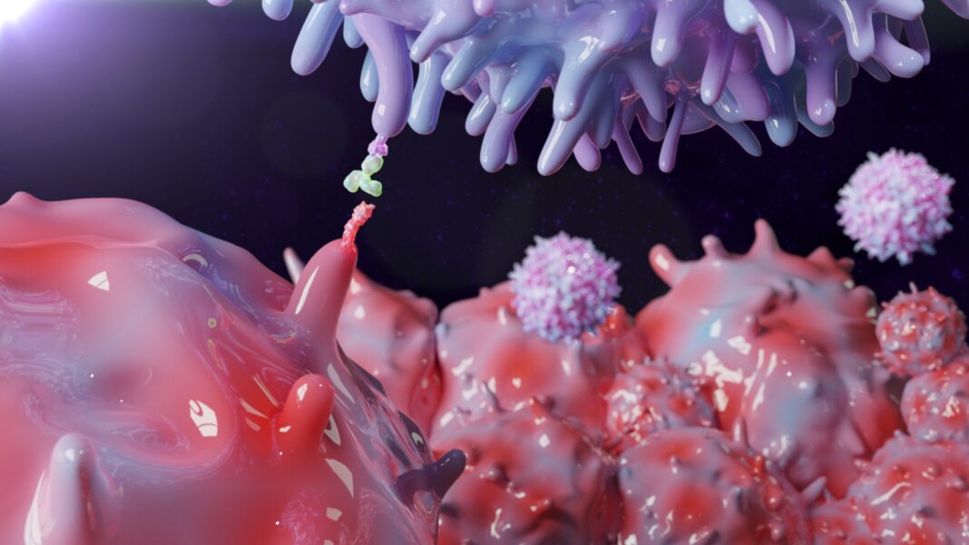
Immune system cells called macrophages play an unexpected role in the complicated connection between obesity and cancer, a Vanderbilt University Medical Center-led research team has discovered. While obesity is a leading risk factor for the progression and metastasis of many cancers, it can in some cases also enhance survival and responses to immune checkpoint blockade therapies, including anti-PD-1 immunotherapy. This is known as the obesity paradox.
The Vanderbilt team’s laboratory experiments and studies in mice with cancer-bearing diet induced obesity (DIO) fed a high-fat diet (HFD) have now shown that obesity increases the frequency of tumor-associated macrophages (TAMs) and induces their expression of the immune checkpoint protein PD-1. The findings provide a mechanistic explanation for how obesity can contribute to both increased cancer risk and enhanced responses to immunotherapy. They may also suggest strategies for improving immunotherapy and for identifying patients who will respond best to such treatments.
The team, headed by Jeffrey Rathmell, PhD, Cornelius Vanderbilt Professor of Immunobiology and director of the Vanderbilt Center for Immunobiology, reported their findings in Nature, in a paper titled “Obesity induces PD-1 on macrophages to suppress anti-tumour immunity.” In their paper the team stated, “These findings provide mechanistic insight into obesity, HFD and immune checkpoint therapies and the role of immune checkpoints in obesity-associated cancers.”
“Obesity is the second leading modifiable risk factor for cancer, behind only smoking, and obese individuals have a greater risk for worse outcomes,” said Rathmell. “But they also can respond better to immunotherapy. How is it that there can be this worse outcome on one hand, but better outcome on another? That’s an interesting question.”
Rathmell and colleagues carried out a series of studies in a high-fat diet (HFD)-fed cancer-bearing diet-induced obese (DIO) mouse model, and in lean mice fed a low-fat diet (LFD), to examine the influence of obesity on cancer and to explore the obesity paradox. “Consistent with obesity promoting cancer growth, our data show that tumours grew more rapidly in mice fed HFD than in those fed LFD,” the team wrote. In addition, while anti-PD-1 therapy attenuated tumor growth in both diet groups of animals, “… a significant decrease in tumor weight occurred online in the HFD-fed mice.”
The studies, led by postdoctoral fellow Jackie Bader, PhD, also showed striking differences between the macrophages isolated from tumors in HFD-fed obese, and from the lean mice. While the protein PD-1 is an immunotherapy target normally thought to act on T cells, the researchers discovered that the macrophages in tumors from obese mice expressed higher levels of PD-1, and that PD-1 acted directly on the macrophages to suppress their function. “The population of PD-1+ macrophages became selectively more abundant with obesity in mouse tumors and inflammation,” the team also pointed out.

“Immunohistochemistry staining on a tumor microarray of more than 100 resected tumors from patients with kidney cancer showed co-localization of both T cell and macrophage markers with PD-1 … with a trending increase in macrophage PD-1 when patients were stratified by BMI,” they wrote. “… immunohistochemistry staining of human endometrial tumor biopsies taken before and after a 10% body weight loss showed significant reduction in the abundance of PD-1-expressing TAMs …” Bader noted. “We were very fortunate to have collaborators that provided us with samples from the same patients before and after weight loss that reinforced the findings from our mouse models.”
Further experiments in the mouse models also showed that anti-PD-1 immunotherapy increased tumor-associated macrophage activity, including their ability to stimulate T cells.
Cancer immunotherapy studies have largely focused on T cells because they are the immune cells that can kill cancer cells, Bader and Rathmell said. But macrophages play important roles in influencing what T cells do. “I’ve always been ‘team macrophage,’” Bader commented. “Macrophages are thought of as being like a garbage truck: They clean up the mess. But they have a huge spectrum of activity to enhance the immune response, and they’re more plastic and manipulatable than other immune cells, which makes them really interesting.”
The presence of more macrophages expressing PD-1 in tumors in an obese setting provides a mechanistic explanation for the obesity paradox, Bader and Rathmell pointed out. Increased PD-1 expression suppresses immune surveillance by macrophages—and subsequently suppresses the killer T cells—allowing tumors to grow (the increased cancer risk of obesity). PD-1 blockade with immunotherapy allows the increased number of PD-1-expressing macrophages to act (the enhanced response to immunotherapy). “These findings identify PD-1 as a metabolic regulator of TAM and reveal a unique PD-1-mediated and macrophage-specific mechanism for immune tumour surveillance and checkpoint blockade,” the investigators wrote. “Taken together, our findings uncover an obesity–cancer connection through induction of PD-1 on macrophages and an alternative mechanism for the obesity paradox of immune checkpoint therapy, in which PD-1 blockade directly enhances TAM-mediated stimulation of anti-tumour T cells.”
Currently, immune checkpoint inhibitors work in only 20-30% of patients. “We clearly want to find ways to make immunotherapies work better, and in the setting [with obesity], they naturally work better,” Rathmell said. “Understanding how these processes are working biologically may give us clues about how to improve immunotherapy in general.”
The findings also suggest that examining levels of PD-1-expressing tumor macrophages may help identify patients who will respond better to immunotherapy, although, as the authors noted, “It remains unclear whether macrophage expression of PD-1 may provide a prognostic indicator for immune checkpoint therapies.” Even so, Rathmell said, “It could be that the greater the proportion of PD-1-expressing macrophages a tumor has, the better the response to immunotherapy will be.”


