Sponsored content brought to you by
![]()
The Emergence of mRNA for Vaccines and Therapy

mRNA vaccines have emerged as perhaps the most promising means to protecting the world’s population from SARS-CoV-2 and future viruses.1 In these vaccines, genetic material coding for a viral protein is encapsulated within a lipid nanoparticle (LNP), which protects it from destructive enzymes and facilitates its delivery into the human body. For the recent Pfizer-BioNTech and Moderna vaccines, the mRNA encodes a harmless piece of the SARS-CoV-2 spike protein (Figure 1). Because the spike protein is unique to the SARS-CoV-2 virus, the human immune system recognizes it as a foreign antigen, thereby resulting in the production of antibodies and T cells against the SARS-CoV-2 virus. This inoculation primes the immune system to better deal with future exposure. Clinical trials have shown that the Pfizer-BioNTech and Moderna mRNA vaccines are highly efficacious at preventing both mild and severe symptoms of COVID-19. Additionally, there have not been any reports of serious safety concerns.2
Beyond their clinical efficacy and safety, mRNA vaccines are advantageous for other reasons. Because mRNA vaccine manufacturing is scalable and cell-free, clinical batches can be manufactured within weeks of discovering an antigen-encoding sequence.3 Furthermore, the development of mRNA vaccines can be much shorter than that of comparable protein-based vaccines because complex cell culture processes are not required for nonclinical research, clinical research, and development studies.4 mRNA vaccine manufacturing (including formulation) is also highly standardized such that a wide variety of vaccines can be rapidly prepared using shared processes and shared facilities.4 The manufacturing firm need only insert or change the coding region of the mRNA to adapt the platform for a new protein or antigen. This means that mRNA vaccines may prove useful in combating new viral variants and to more effectively control seasonal influenza strains.5
With the emergency authorization of mRNA COVID-19 vaccines, there is more momentum than ever for mRNA to be used as a modality for other therapeutic approaches, like treating rare genetic disorders in both gene replacement and gene editing capacities. In one case, mRNA encoding an enzyme implicated in lysosomal storage disorders is being studied.6 And in another example, mRNA for Cas proteins is being paired with guide RNA to provide in vivo CRISPR gene editing.7 There is a significant wave of progress at hand, and it has been unlocked by advances in lipid nanoparticles as well as pseudouridine and methyl-pseudouridine residues that have helped unlock high levels of intracellular translation.3
As this new form of biotechnology makes a leap of progress, so too do the analytical approaches supporting its development. From an analytical perspective, LNP-encapsulated mRNA molecules need to be characterized and QC tested to ensure safety and efficacy. Modern techniques including liquid chromatography (LC), mass spectrometry (MS), and differential calorimetry (DSC) are therefore being applied to analyze important lipid and mRNA attributes. Here we will touch on three key analytical strategies for ensuring that safe and efficacious encapsulated mRNA drugs are developed and manufactured:
- LNP purity and composition by LC
- Differential scanning calorimetry for LNP formulation stability
- mRNA Analysis by LC and LC-MS methods
Lipid Nanoparticles Are the Key to Delivery
In and of themselves, LNPs represent a breakthrough technology. LNPs are the preferred delivery vessel for mRNA therapies and vaccines. For vaccines, they have the added benefit of stimulating the innate immune system without inducing immunogenicity. LNPs are engineered with a specific pKa and size which facilitates optimal protein expression and immunogenicity. These nanoparticles are approximately 50 to 100 nm in diameter and are created through the mixing of the four different types of lipids (Figure 2).8 Each component is combined at a specific stoichiometric ratio, and nanoparticle formation is initiated by the controlled mixing of aqueous mRNA with an alcohol solution of the lipids. These lipids include an ionizable, cationic lipid (such as MC3), a zwitterionic phospholipid (such as DSPC), cholesterol, and a pegylated lipid (such as PEG-DMG).9,10

There is an important role for each lipid to play. The ionizable cationic lipid is electrostatically attracted to the negatively charged backbone of the mRNA, and it initiates the formation of an inverted micelle. Meanwhile, the cholesterol and zwitterionic phospholipid serve as bulking agent, and the pegylated lipid stabilizes an energetically favorable surface layer. Progress on lipid chemistry continues, but these lipids have served as the formulation for patisiran, a transthyretin-directed small interfering RNA,11 and for some of Moderna’s earliest clinical studies.12 Novel lipids and new mixtures are in development and can now even be found in the Pfizer-BioNTech COVID-19 vaccine, but the general architecture of the LNP is likely to persist regardless of new developments in lipid technology, such as the use of alternative cationic lipids.8
Chromatography Checks on Purity and Lipid Composition
As one might predict, the purity of the lipids used in the LNP matters, and this is an important attribute to check during preclinical, clinical, and product stages of a new vaccine or therapy. Where needed, lab-scale chromatographic purification can be applied to further purify lipid raw materials. Interestingly, there is an opportunity to consider supercritical fluid chromatography for this purpose and to create a green chemistry purification scheme with carbon dioxide as the only chemical waste, not excess volumes of organic solvents.13 Preparative-scale LC with up to 50-mm internal diameter columns and up to 150-mL/min flow rates can also be employed.
With the lipid reagents in hand, purity can be assayed by gas chromatography (GC) and MS. When these techniques are used together, in GC-MS analysis, it is often possible to comprehensively identify and quantitate lipid impurities. LC paired with MS offers potential advantages for impurity identification as well. LC instrumentation can be applied with ESI-MS detection, using method insights first developed for lipidomic studies.14 Impurities corresponding to degradation, oxidation, and synthesis side reactions can be identified according to accurate mass information, and structural information can be deciphered from the application of various types of gas-phase fragmentation and subsequent MS/MS analysis.

Yet another way that LC is being applied to the analysis of lipids is in the determination of each lipid’s relative abundance in a formulated LNP-encapsulated mRNA. In one important analysis, the lipids are chromatographically separated using a form of reversed-phase chromatography and then detected by evaporative light-scattering detection and potentially even MS. This assay has been used through stages of development as well batch release. Preliminary work suggests that a Waters™ ACQUITY™ PREMIER LC system, with its low-adsorption hybrid surfaces,15 might prove useful to establish more robust relative quantitation on account of it facilitating improved recovery of phospholipids (Figure 3).16 Initial work on these separations has also shown that PREMIER columns packed with a charged surface hybrid stationary phase provide advantageous selectivity and peak shape with the low ionic strength formic acid/formate-based mobile phases preferred for evaporative light-scattering detection.17 With these LC approaches being more widely adopted, it is foreseeable that MS detection will also become more routine. It might soon be possible to implement regulatory-compliant MS along with new peak detection for a comprehensive analytical approach akin to peptide-level multi-attribute monitoring on monoclonal antibodies.18

DSC for LNP Formulation Studies
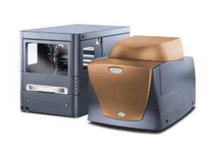
DSC is a thermal analysis technique that measures changes as a function of temperature by either heating and/or cooling a sample to induce a transition (Figure 4). For complex lipids, such as LNPs, the transitions of interest include phase changes, confirmational changes in their packing, and melting temperatures. These events can be observed in both the raw material for investigation of contaminants and on the colloidally dispersed state for general characterization. A colloid solution is formed through the self-assembly of LNPs. This is a spontaneous event, and the larger complex is held together by noncovalent interactions where these weak intermolecular forces allow structural changes as the complex moves toward equilibrium.19 The state initially adopted is typically a “metastable state.” Upon reheating the sample, they change their configuration into a more stable form that persists upon additional heat cycles in a DSC. This can be problematic especially when considering the impact of longer-term storage if the metastable state is the preferred configuration. Due to a structure-activity relationship (SAR) of an LNP, a different form could have deleterious effects as it will preferentially change cellular or organ uptake.19
An ideally formulated LNP contains multiple components, each responsible for a different attribute and each impacting the higher-order structure (HOS). One example is the addition of cholesterol added to enhance LNP stability and promote membrane fusion.20 In the DSC, the addition of cholesterol presents itself as a broad endotherm, and it often decreases the enthalpy of the transition without changing the Tmax. Other lipid combinations will impact the transition temperatures, for example, the increase of the Tmax of a complex event versus its individual components is attributed to the formation of strong electrostatic interactions between the positively charged ammonium ion from the cationic lipid and the zwitterionic phospholipid.21 The other element that will change the phase transition temperature is the addition of the mRNA. The structure will be perturbed upon the addition of the highly charged molecule, especially when considering the involvement of so many forces, like electrostatic interactions, hydrophobic interactions, and van der Waals forces. The composition of the LNP, which is related to function and potency, can be correlated to HOS alterations and changes in transition temperatures through the use of a DSC instrument.
mRNA Analysis by LC and LC-MS
The messenger RNA that is manufactured for a vaccine or therapy is produced through a cell-free process known as in vitro transcription (IVT) synthesis, which produces millions of copies of the molecule from an initial plasmid DNA template. After mRNA has been IVT-synthesized, it is processed by enzymes that add a guanine nucleotide to its 5’ end (5’ cap), and a chain of several adenine nucleotides to its 3’ end. These post-transcriptional modifications are considered critical quality attributes as they help protect the IVT-synthesized mRNA from degradation and have an impact on translation efficiency as well.22
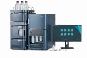
In the development of mRNA-based therapeutics and vaccines, LC and LC-MS workflows are used to ensure proper addition of the 5’ cap and polyA tail, as well as to measure capping efficiency and confirm 5’ cap identity and polyA tail length. For the determination of 5’ capping efficiency and identity, Beverly and co-workers from the Novartis Institutes of Biomedical Research devised a method utilizing specific RNase H probes to cleave off 50 bases from the 5’ end of the mRNA. With a biotin-streptavidin-enriched sample, they were then able to perform LC-MS analysis of the cleaved fragments.23 In another paper, Beverly and colleagues combined an RNase digestion with polydT affinity enrichment to collect polyA tail oligonucleotides from an mRNA, and they then subjected the sample to LC-MS analysis.24 In this more recent work, MS detection was shown to be capable of directly measuring multiple oligonucleotide sequences with single-nucleotide resolution. Consequently, it was demonstrated that the approach was suited to the study of mRNA poly A tail length. Permutations of these mRNA fragment analyses are being considered for both characterization and QC testing. Advances in analytical technology are making it possible to robustly deploy these assays across the world. New mobile-phase systems based on hexylammonium acetate and diisopropylethylamine afford high peak capacity along with unprecedented MS sensitivity.25,26 Also, because it was designed for regulatory-compliant work, the Waters BioAccord LC-MS system makes it possible to carry through the use of one instrument from development and preclinical testing to batch release QC testing (Figure 5). With respect to chromatography, the recent commercialization of PREMIER Oligonucleotide BEH C18 columns has proven to be fortuitous. PREMIER columns, with their hybrid surface column hardware, alleviate the well- known concerns of oligonucleotides adsorbing to metal hardware, which means that right-the-first time results can be achieved without concerns over column-to-column reproducibility and without needing to suffer through a process of conditioning new columns with repeat sample injections.27
In addition to these examples of mRNA fragment analysis, some researchers are exploring an orthogonal method for confirming mRNA identity. Hua and co-workers have explored a so-called oligo-mapping approach that is somewhat analogous to peptide mapping methods for protein-based therapeutics. Here too, the mRNA is digested, and the resulting small oligonucleotide fragments are separated via ion pairing reversed-phase LC to generate an oligonucleotide map that can serve as a fingerprint to verify mRNA ID and sequence.28 Lastly, there is interest in studying the mRNA at its intact level, which is typically 2,000 to 5,000+ nucleotides long. With the pace of advances in MS, it is quite likely that we are on the cusp of such an analytical approach. If not by MS, an analyst might choose to apply size-exclusion chromatography or an ion-exchange separation29 for an intact level analysis of the molecule.
Learn more Waters.com/CGT
References
1. Alkandari, D.; Herbert, J. A.; Alkhalaf, M. A.; Yates, C.; Panagiotou, S., SARS-CoV-2 vaccines: fast track versus efficacy. The Lancet Microbe 2021.
2. Maragakis, L. L.; Kelen, G. D. Is the COVID-19 Vaccine Safe?
3. Jackson, N. A. C.; Kester, K. E.; Casimiro, D.; Gurunathan, S.; DeRosa, F., The promise of mRNA vaccines: a biotech and industrial perspective. NPJ Vaccines 2020, 5 (1), 11.
4. The Advantages of mRNA Vaccines
5. Einstein, M. For mRNA vaccines, COVID-19 is just the beginning.
6. Zhu, X.; Yin, L.; Theisen, M.; Zhuo, J.; Siddiqui, S.; Levy, B.; Presnyak, V.; Frassetto, A.; Milton, J.; Salerno, T.; Benenato, K. E.; Milano, J.; Lynn, A.; Sabnis, S.; Burke, K.; Besin, G.; Lukacs, C. M.; Guey, L. T.; Finn, P. F.; Martini, P. G. V., Systemic mRNA Therapy for the Treatment of Fabry Disease: Preclinical Studies in Wild-Type Mice, Fabry Mouse Model, and Wild-Type Non-human Primates. Am J Hum Genet 2019, 104 (4), 625-637.
7. Idrus, A. A. Using gene editing to keep cholesterol down and heart attacks at bay.
8. Buschmann, M. D.; Carrasco, M. J.; Alishetty, S.; Paige, M.; Alameh, M. G.; Weissman, D., Nanomaterial Delivery Systems for mRNA Vaccines. Vaccines (Basel) 2021, 9 (1).
9. Hassett, K. J.; Benenato, K. E.; Jacquinet, E.; Lee, A.; Woods, A.; Yuzhakov, O.; Himansu, S.; Deterling, J.; Geilich, B. M.; Ketova, T.; Mihai, C.; Lynn, A.; McFadyen, I.; Moore, M. J.; Senn, J. J.; Stanton, M. G.; Almarsson, O.; Ciaramella, G.; Brito, L. A., Optimization of Lipid Nanoparticles for Intramuscular Administration of mRNA Vaccines. Mol Ther Nucleic Acids 2019, 15, 1-11.
10. Wadhwa, A.; Aljabbari, A.; Lokras, A.; Foged, C.; Thakur, A., Opportunities and Challenges in the Delivery of mRNA-based Vaccines. Pharmaceutics 2020, 12 (2).
11. Adams, D.; Gonzalez-Duarte, A.; O’Riordan, W. D.; Yang, C. C.; Ueda, M.; Kristen, A. V.; Tournev, I.; Schmidt, H. H.; Coelho, T.; Berk, J. L.; Lin, K. P.; Vita, G.; Attarian, S.; Plante-Bordeneuve, V.; Mezei, M. M.; Campistol, J. M.; Buades, J.; Brannagan, T. H., 3rd; Kim, B. J.; Oh, J.; Parman, Y.; Sekijima, Y.; Hawkins, P. N.; Solomon, S. D.; Polydefkis, M.; Dyck, P. J.; Gandhi, P. J.; Goyal, S.; Chen, J.; Strahs, A. L.; Nochur, S. V.; Sweetser, M. T.; Garg, P. P.; Vaishnaw, A. K.; Gollob, J. A.; Suhr, O. B., Patisiran, an RNAi Therapeutic, for Hereditary Transthyretin Amyloidosis. N Engl J Med 2018, 379 (1), 11-21.
12. Bahl, K.; Senn, J. J.; Yuzhakov, O.; Bulychev, A.; Brito, L. A.; Hassett, K. J.; Laska, M. E.; Smith, M.; Almarsson, O.; Thompson, J.; Ribeiro, A. M.; Watson, M.; Zaks, T.; Ciaramella, G., Preclinical and Clinical Demonstration of Immunogenicity by mRNA Vaccines against H10N8 and H7N9 Influenza Viruses. Mol Ther 2017, 25 (6), 1316-1327.
13. Beginner’s Guide to Preparative SFC. Waters 2017.
14. Isaac, G.; McDonald, S.; Astarita, G., Lipid Separation using UPLC with Charged Surface Hybrid Technology. Waters Application Note 720004107 2011.
15. T. H. Walter, M. T., J. Simeone, P. Rainville, A. V. Patel, M. A. Lauber, J. Kellett, M. DeLano, K. Brennan, C. Boissel, R. Birdsall, K. BertheletteWaters Corporation, Milford, MA, USA, Low Adsorption UPLC Systems and Columns Based on MaxPeak High Performance Surfaces: The ACQUITY PREMIER Solution. Waters White Paper 720007128 2021.
16. Isaac, G.; Plumb, R., ACQUITY PREMIER LC Technology Significantly Improves Sensitivity, Peak Shape, and Recovery for Phosphorylated and Carboxylate Lipids Waters Application Note 720007092 2021.
17. Iraneta, P. c.; Kevin D. wyndham; Mccabe, D. R.; walter, T. H., Charged surface Hybrid (csH) technology and its Use in liquid chromatography. Waters White Paper 20003929EN 2011.
18. Ranbaduge, N.; Yu, Y. Q., A Streamlined Compliant Ready Workflow for Peptide-Based Multi-Attribute Method (MAM). Waters Application Note 720007094 2021.
19. Kim, J.; Eygeris, Y.; Gupta, M.; Sahay, G., Self-assembled mRNA vaccines. Adv Drug Deliv Rev 2021, 170, 83-112.
20. Kauffman, K. J.; Dorkin, J. R.; Yang, J. H.; Heartlein, M. W.; DeRosa, F.; Mir, F. F.; Fenton, O. S.; Anderson, D. G., Optimization of Lipid Nanoparticle Formulations for mRNA Deliver in Vivo with fractional factorial and definitive screening designs. Nano Lett. 2015, (15), 7300-7306.
21. Ziller, A.; Nogueira, S. S.; Huhn, E.; Funari, S. S.; Brezesinski, G.; Hartmann, H.; Sahin, U.; Haas, H.; Langguth, P., Incorporation of mRNA in Lamellar Lipid Matrices for Parenteral Administration. Mol Pharm 2018, 15 (2), 642-651.
22. Pardi, N.; Hogan, M. J.; Porter, F. W.; Weissman, D., mRNA vaccines – a new era in vaccinology. Nat Rev Drug Discov 2018, 17 (4), 261-279.
23. Beverly, M.; Dell, A.; Parmar, P.; Houghton, L., Label-free analysis of mRNA capping efficiency using RNase H probes and LC-MS. Analytical and Bioanalytical Chemistry 2016, (408), 5021–5030
24. Beverly, M.; Hagen, C.; Slack, O., Poly A tail length analysis of in vitro transcribed mRNA by LC-MS. Anal Bioanal Chem 2018, 410 (6), 1667-1677.
25. Gong, L.; McCullagh, J. S., Comparing ion-pairing reagents and sample dissolution solvents for ion-pairing reversed-phase liquid chromatography/electrospray ionization mass spectrometry analysis of oligonucleotides. Rapid Commun Mass Spectrom 2014, 28 (4), 339-50.
26. McCarthy, S.; Gilar, M., Hexylammonium Acetate as an Ion-pairing Agent for IP-RP LC Analysis of Oligonucleotides. Waters Application Note 720003361 November 2016.
27. Brennan, K.; Trudeau, M.; Rainville, P. D., Utilization of the ACQUITY PREMIER System and Column for Improved Oligonucleotide Bioanalytical Chromatographic Performance. Waters Application Note 720007119 2011.
28. Jiang, T.; Yu, N.; Kim, J.; Murgo, J. R.; Kissai, M.; Ravichandran, K.; Miracco, E. J.; Presnyak, V.; Hua, S., Oligonucleotide Sequence Mapping of Large Therapeutic mRNAs via Parallel Ribonuclease Digestions and LC-MS/MS. Anal Chem 2019, 91 (13), 8500-8506.
29. Kanavarioti, A., HPLC methods for purity evaluation of man-made single-stranded RNAs. Sci Rep 2019, 9 (1), 1019.