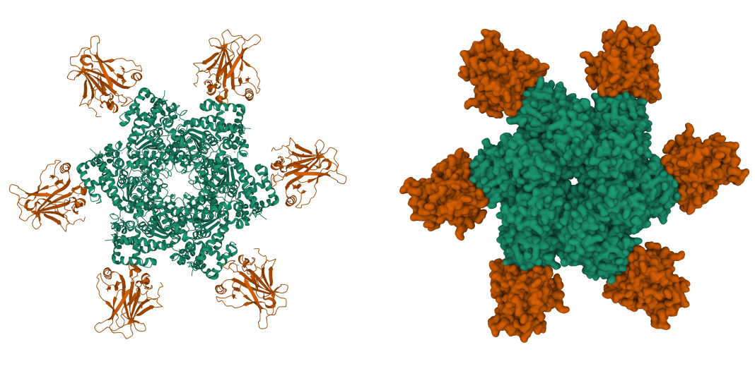
Guanine quadruplexes (G-quadruplexes or G4s) are unusual nucleic acid structures with distinct biological and chemical functions. They have occupied significant attention in the nucleic acid world since they were first identified in 1962 as the basis for the aggregation of 5’-guanosine monophosphate molecules.
Guanine-rich sequences in DNA or RNA have the unique ability to self-assemble into planar arrangements known as G-quartets. In the presence of metal cations (such as K+ or Na+), G-quartets can stack atop each other and fold to form stable G4 structures with different numbers of associated strands (molecularity) and surface features (topology).

Putative Quadruplex Sequences (PQS) are highly abundant in key regions of human genes such as telomeres and promoters and are believed to play an essential role in replication and gene regulation.1 In addition, G4s are vital elements in regulating the lifecycle of RNA viruses, including human immunodeficiency virus (HIV),2 Epstein-Barr viruses (EBV),3 Hepatitis C virus (HCV),4 and SARS-CoV-2.5 Many G4-stabilizing compounds have shown potent antiviral activity and therapeutic effects for diseases like cancer.6
While the chemical and physical properties of G4 structures are fascinating on their own, they provide the fingerprint for distinguishing between quadruplex and duplex nucleic acid structures. If you are studying nucleic acid structures in your lab, you may apply several methods to identify and characterize G4 structures and confirm their formation in your samples. These methods can be based on molecular biology assays or biophysical techniques. This article covers three standard biophysical techniques to detect G4s.
Fluorescence/absorbance turn-on assays
Many fluorogenic compounds and dyes bind and stabilize G-quadruplexes. Upon binding, the complex exhibits altered physical properties, changing its fluorescence or absorbance intensity. For example, porphyrins such as heme bind G4s strongly and dramatically change the UV-visible spectrum of free heme. These changes are unique and specific to G4 structures and don’t occur in duplexes or single-stranded DNA or RNA.7
This method is widely used to detect G-quadruplexes because it is simple and cheap, but it is limited by the availability, sensitivity, and binding affinity of the fluorogenic compounds or dyes. Also, some compounds may not bind to all G4s topologies.
Circular dichroism (CD) spectroscopy
G4s produce a distinctive CD spectrum because the stacked G-quartet units are rotated with respect to each other and can absorb circular polarized light differently. Depending on the direction of the phosphate backbone and glycosidic bond angles as well as the number of strands associated, G4s can adopt various surface characteristics and can be classified into “parallel,” “hybrid,” and “antiparallel”.

Each of these conformations can have a different CD signal. Here are example of some well-known G4 sequences that adopt these different molecular and surface features and their corresponding CD signatures.The CD spectrum of the parallel d(TGGGGT)4 is characterized by a positive peak at 260 nm and a negative peak at 240 nm. For hybrids like d[T2G3(T2AG3)3A], the CD spectrum has an additional positive peak at 290 nm. In contrast, the bimolecular antiparallel d(G4T4G4)2 quadruplex is characterized by a CD spectrum with a positive peak in the region between 290 and 300 nm and a negative peak between 240 and 260 nm. Irrespective of the number of stacked G-quartets, these observations have been generally adopted to explain the difference between parallel, hybrid, and antipatallel G-quaduplexes.8

1H nuclear magnetic resonance (NMR)
In contrast to Watson-Crick hydrogen bonding, which involves N1 and N3 of the heterocyclic rings, the G-quartet array forms a different type of hydrogen bonding involving N7. This bonding system is known as Hoogsteen hydrogen-bonding, and it allows the purines to be in an unusual conformation and permits the detection of the imino protons of guanine by 1H NMR. Hoogsteen hydrogen bonds in G-quartets display chemical shifts at 10.2 – 11.6 parts per million (ppm) which are considered the hallmark of the G-quadruplex structures.10
The method you adopt to characterize G4s in your sample will depend on the availability of spectroscopic reagents and instruments, and the nature of the project.
References
- Rhodes D, Lipps HJ. G-quadruplexes and their regulatory roles in biology. Nucleic Acids Res. 2015;43(18):8627-8637. doi:10.1093/nar/gkv862
2. Piekna-Przybylska D, Sullivan MA, Sharma G, Bambara RA. U3 Region in the HIV-1 Genome Adopts a G-Quadruplex Structure in Its RNA and DNA Sequence. Biochemistry. 2014;53(16):2581-2593. doi:10.1021/bi4016692
3. Murat P, Zhong J, Lekieffre L, et al. G-quadruplexes regulate Epstein-Barr virus–encoded nuclear antigen 1 mRNA translation. Nat Chem Biol. 2014;10(5):358-364. doi:10.1038/nchembio.1479
4. Wang SR, Min YQ, Wang JQ, et al. A highly conserved G-rich consensus sequence in hepatitis C virus core gene represents a new anti–hepatitis C target. Sci Adv. 2(4):e1501535. doi:10.1126/sciadv.1501535
5. Zhao C, Qin G, Niu J, et al. Targeting RNA G-Quadruplex in SARS-CoV-2: A Promising Therapeutic Target for COVID-19? Angew Chem Int Ed. 2021;60(1):432-438. doi:10.1002/anie.202011419
6. Kosiol N, Juranek S, Brossart P, Heine A, Paeschke K. G-quadruplexes: a promising target for cancer therapy. Mol Cancer. 2021;20(1):40. doi:10.1186/s12943-021-01328-4
7. Shumayrikh N, Sen D. Heme•G-Quadruplex DNAzymes: Conditions for Maximizing Their Peroxidase Activity. In: Yang D, Lin C, eds. G-Quadruplex Nucleic Acids: Methods and Protocols. Methods in Molecular Biology. Springer; 2019:357-368. doi:10.1007/978-1-4939-9666-7_22
8. Carvalho J, Queiroz JA, Cruz C. Circular Dichroism of G-Quadruplex: a Laboratory Experiment for the Study of Topology and Ligand Binding. ACS Publications. doi:10.1021/acs.jchemed.7b00160
9. Randazzo A, Spada GP, da Silva MW. Circular Dichroism of Quadruplex Structures. In: Chaires JB, Graves D, eds. Quadruplex Nucleic Acids. Topics in Current Chemistry. Springer; 2013:67-86. doi:10.1007/128_2012_331
10. Binas O, Bessi I, Schwalbe H. Structure Validation of G‐Rich RNAs in Noncoding Regions of the Human Genome. Chembiochem. 2020;21(11):1656-1663. doi:10.1002/cbic.201900696


