October 1, 2014 (Vol. 34, No. 17)
Utilizing Approaches Based on Reversed-Phase and Gel Filtration Chromatography
Protein drugs feature significant advantages over small molecule therapeutics, including low toxicity, high specificity, and development speed.
However, the quality control requirements are significantly more complex in that a variety of QC tests are required to verify the composition of the protein as well as to quantitate any minor impurities due to post-translational modification of the protein of interest. Quantitating these aberrant proteins is especially important as such modifications are usually the proximal cause of potential life-threatening neutralizing antibodies to the therapeutic.
While most sequence-specific modifications are best identified by peptide mapping of the proteolytic digest of the protein, other global modifications like folding irregularities and protein aggregation are best identified by intact protein analysis (e.g., reversed phases, ion-exchange chromatography, and gel filtration chromatography), which allows one to get some information about the entire folded protein rather than just a specific amino acid.
In this article, two of those three methods—intact reversed-phase chromatography and gel filtration chromatography—are discussed. Each technique has its unique challenges and strategies for optimization. Especially with some of the new UHPLC column and instrument technologies, there are some specific ways of optimizing both methods to generate the desired quantitation of post-translational modifications in a reliable and reproducible manner.
Materials and Methods
Protein standards and all buffer additives were purchased from Sigma Chemical. Mobile-phase solvents were purchased from EMD Millipore. The columns used for reversed-phase analysis of intact proteins were the core-shell Aeris Widepore 3.6 µm C4, XB-C8, and XB-C18 (150 × 4.6 mm), and the column used for gel filtration analysis was a Yarra 3 µm GFC3000 (300 × 7.8 mm dimension; Phenomenex).
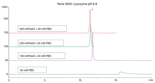
All samples were run on the Agilent 1260 HPLC with solvent degasser, auto sampler, column oven, and UV detector. Data was collected using Agilent’s Chemstation software. For reversed-phase analysis the aqueous mobile phase used 0.1% TFA. The organic mobile phase used was acetonitrile with 0.085% TFA. For gel filtration analysis the mobile phase used was 100 mM sodium phosphate, with and without sodium chloride (Figure 2) pH 6.8.

Figure 2. Gel filtration chromatography of lysozyme on a Yarra SEC-3000 column (300 x 7.8 mm) using different mobile-phase conditions of increasing salt. Note that as salt concentration increased, peak shape and recovery for lysozyme improved. pH, salt concentration, and organic additives can all influence secondary interactions between GFC stationary phase and protein analytes.
Reversed-Phase Analysis: Method Optimization
Reversed-phase analysis of intact proteins often is useful for quantitating changes in protein folding as well as in some single amino acid post-translational modifications. Key for any such method is obtaining good recovery of proteins as well as resolution of modified proteins that are chemically similar to the protein therapeutic.
Protein recovery using core-shell UHPLC protein columns is not as problematic as with older fully porous 300 Å columns, but can be improved by increasing column temperature, which also can help improve peak shape and resolution. Operating column temperature up to 90°C can be used to improve recovery and peak shape.
As temperature increases, peak widths of the major protein peaks decrease and absolute retention of each peak is reduced with increasing temperature. In extreme cases with very large or hydrophobic proteins, the addition of isopropanol in the organic mobile phase can improve protein recovery (data not shown).
Additional steps for improving resolution of intact protein revolve around modifying the gradient slope of the protein separation as well as evaluating the stationary phase used (C4, C8, or C18) for the separation. As with other reversed-phase gradient separation methods, shallowing the organic gradient slope will usually improve resolution of proteins but at a cost of longer run times and wider protein peaks.
In developing a gradient method on core-shell protein columns, one must also reduce initial organic concentration as such columns are far less retentive than their fully porous predecessors. Stationary-phase chemistry can also have an impact on the resolution of proteins. Contrary to previous paradigms that used a 300 Å C4 for proteins and a 300 Å C18 column for peptides, core-shell columns of all chemistries can be used to optimize a separation for both proteins and peptides.
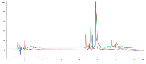
An example of selectivity differences is shown in Figure 1 where the same Ig-G antibody was run on three different protein widepore core-shell columns. Note the changes in resolution and selectivity of the major peaks in the chromatogram based on the stationary phase used.
Despite reports by others in the past, column chemistry can make a difference in reversed-phase protein separations.

Figure 1. Protein selectivity of Ig-G1 using different stationary phases. In this example, a human Ig-G1 was run on three different Aeris Widepore 3.6 µm columns (C4, XB-C8, and XB-C18; all 150 x 4.6 mm) using a rapid gradient separation (20–60% B in 14 minutes). Note the change in resolution of the major components based on stationary phase used. Stationary-phase chemistry can be screened to find the optimal separation for a particular protein.
Gel Filtration Analysis: Method Optimization
Gel filtration chromatography (GFC) is commonly used when analyzing therapeutic proteins to determine the amount of protein aggregate present in a sample. GFC is thought by many to be the simplest form of chromatography, with no method optimization required beyond picking the pore size of the GFC column based on molecular weight ranges, but in reality that is not the case.
GFC columns are bonded with a polar stationary phase to minimize interactions between the protein and the column such that separation is only based on a protein’s ability to permeate the pores of the media leading to increasing protein retention inverse to a protein’s size in solution (which is closely related to the molecular weight of a protein).
In an ideal situation that would be the case. Unfortunately some secondary interactions, both hydrophobic and ionic, between the stationary phase and protein analytes do occur and mobile-phase optimization of GFC methods revolves around minimizing the strongest interaction in separation based on whether it is ionic or hydrophobic.
An example of suppressing ionic interactions using increasing salt concentrations is shown in Figure 2 where a basic protein (lysozyme) is run under increasing salt concentrations. At low salt concentrations the lysozyme peaks elute anomalously with poor peak shape and recovery. As salt concentration of the mobile phase increases, peak shape and recovery improves.
While this would tend to lead one to use high salt concentrations for all GFC separations, it is a balance between interactions. With increasing salt concentrations, hydrophobic interactions increase between protein analytes and the stationary phase leading to recovery decreases for hydrophobic proteins (data not shown).
Another factor that influences resolution of GFC (besides the column used) is the flow rate used for a separation. As is shown in Figure 3, when the efficiency of a protein mixture run on a GFC column is plotted out versus flow rate one can see that large proteins have increasingly better resolution by GFC at lower flow rates (versus small proteins, which perform better at high linear velocities). Similar to reversed-phase protein analysis it is all a matter of the time that one wants to spend on a separation versus the resolution one wishes to achieve.
Intact protein analysis using various chromatography techniques is a critical part of the portfolio of protein characterization experiments that must be performed on a protein therapeutic in a product development or lot-release situation. While such methods are thought to have limited method-development possibilities, data shown here demonstrates that one actually has several method-development options when working with modern chromatography columns.

Figure 3. Gel filtration chromatography of a protein standard at different flow rates. The plate height (inverse of efficiency) of a protein standard on a Yarra SEC-3000 is plotted at different flow rates. Note that for many large proteins, the plate height decreases (increasing efficiency) with lower flow rate, provided that one is willing to experience long run times.
Michael McGinley ([email protected]) is senior bioseparations product
manager, Lauren Mitre is a QA scientist, and Michael Klein is HPLC and UHPLC brand manager at Phenomenex.


