November 1, 2014 (Vol. 34, No. 19)
A Simple Touchscreen Fluorescence Cell Imager Improves Workflow for Routine Applications
Whether it’s for gaining a better understanding of how cells work, studying the effects of drugs or toxins on cells, or developing new and better medicines, cell culture researchers routinely use fluorescence imaging. Fluorescence microscopy is the most widespread technique for studying live and fixed cells, and is integral to nearly every cell culture application.
Epifluorescence microscopes have become the instrument of choice for routine fluorescence imaging since they were developed in the 1960s. Despite being widely used, epifluorescence microscopes have characteristic issues that have frustrated researchers since their conception. Fluorescence imaging requires specialized equipment and knowledge often prompting researchers to search out knowledgeable experts in their lab.
Imaging has come a long way since the mid-20th century. However, there is still room for improvement. The usability of epifluorescence microscopes can be disruptive to the workflow of scientists working on cell culture research.
Sizing Up Challenges of Traditional Fluorescence Microscopy
The fluorescence microscopy setup in most labs is cumbersome. The microscopes are often located in a darkroom away from the researchers’ main workspace. Even the system itself may be a convoluted tangle of cables connecting an inverted benchtop fluorescence microscope to image acquisition, visualization, analysis, and storage equipment.
Fluorescence microscope systems are so complicated that Checo Rorie, Ph.D., of North Carolina Agricultural and Technical State University says there are certain students that still can’t master the microscopes no matter how hard they try. Often, students must put in hours of practice before they’re able to produce crisp images. Researchers who are able to master fluorescence skills default into an “in-house microscopy expert” position. Instead of being able to focus on their own work uninterrupted, their colleagues seek them for help navigating epifluorescence instruments. During periods of heavy use, these experts can become overburdened and limit the lab’s productivity.
Systems are also kept in a darkroom since ambient light can cloud an image collected from a fluorescent microscope. In many research institutions space is limited so these types of specialized facilities are usually shared between research groups, which can lead to scheduling backups. Demand for instrument time can run very high, limiting how quickly work can get done for each group. Using instruments can get even more complicated when another group forgets to reset the settings.
There is another hidden cost to using fluorescence microscopes: the darkroom. Researchers may need to travel from their lab to work in the darkroom. Frustration of lost time builds up as the researcher shuttles back and forth between the lab and the darkroom. Some scientists report losing up to 30 minutes from travel and time spent warming up the microscope. Those that have to work for an extended period of time in a darkroom, alone and away from people, often find it depressing.
An Alternative to Streamline Cell Culture Applications
These challenges highlight an unmet need for a device that eliminates the drawbacks involved in today’s cell culture workflow. Bio-Rad Laboratories has developed an innovative solution that requires no training, renders the darkroom unnecessary, and fits easily on the workbench and in the budget of an average cell culture laboratory.
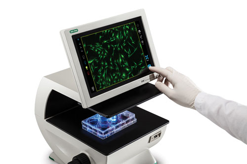
The ZOE™ fluorescence cell imager streamlines the cell culture workflow. ZOE is a simple cell imager that combines the functions of a traditional brightfield microscope with red, green, and blue fluorescent channels (Figure 1). ZOE’s intuitive touchscreen user interface is easy to use and takes little time to learn how to operate (Figure 2). Practically anyone can turn the imager on and start using it.

Figure 1. The ZOE fluorescence cell imager software allows researchers to edit each image as well as overlay and merge into multicolor images. Left: User interface showing Live mode with Edit menu in the brightfield channel. Right: HEK 293 cells transfected with enhanced green fluorescent protein and red fluorescent protein.
ZOE offers additional advantages over traditional fluorescence microscopes. Researchers can keep the fluorescent imager in their lab on their benchtop. ZOE’s display screen makes it easy to observe a specimen through a lens while also fostering collaboration and discussion as multiple people can view the specimen at the same time. Additionally, the light shield allows researchers to image a sample without turning off the lights.
ZOE’s LED light sources minimize the photobleaching effect because their intensity can be tuned to fit the researcher’s needs. LEDs also deliver a more constant source of light and can last tens of thousands of hours. In contrast, the illumination provided by mercury arc lamps in fluorescence microscopes degrades slowly over time and the lamps must be replaced after 300 hours. Another advantage is a built-in camera that can take pictures of the specimens for analysis or documentation.
Many researchers use a confocal microscope to capture publication quality images; however, confocal microscopes require a significant amount of prep work. Washing cells and staining them with antibodies can take multiple days. If something goes wrong during the process, they may have wasted the time and effort that went into using the confocal microscope.
ZOE can be conveniently located in the lab allowing researchers to easily optimize or check experimental conditions. They can quickly check to see if the staining worked. Once researchers are reasonably assured of their results, they can make the decision to invest the time and effort needed to obtain publication quality images on a confocal microscope.
With the arrival of the ZOE fluorescent cell imager, cell culture labs are going to become a little bit brighter. Cell culture researchers can finally ditch the darkroom for good and say good-bye to the inconvenience, frustration, and wasted time of using a fluorescence microscope.

Figure 2. Bio-Rad’s ZOE fluorescence cell imager uses an intuitive high-resolution LCD touchscreen interface, which is compatible with gloved hands.
Veronika Kortisova-Descamps ([email protected]) is a product manager at Bio-Rad Laboratories.


