May 1, 2016 (Vol. 36, No. 9)
Tools for PD-1 and TIGIT Immune Checkpoint Antibody Development
Therapeutic antibodies designed to block immune checkpoint receptors or activate immunostimulatory receptors are a promising strategy to treat cancer and function by modulating a patient’s own immune system. Co-engagement of multiple immune receptors on chronically activated T cells, known as combination immunotherapy, can potentially elicit better therapeutic outcomes through a combinatorial effect compared with engagement of a single receptor.
The results of numerous clinical trials support the strategy of combination immunotherapy. For example, PD-1/PD-L1 blocking antibodies, which have shown positive clinical results, are now being evaluated in combination with antibody drugs that target additional immune checkpoint receptors.
TIGIT is a target immune checkpoint receptor that is expressed on subsets of activated T cells and natural killer (NK) cells. The ligand for TIGIT is CD155 (also called poliovirus receptor) that is, interestingly, also a ligand for CD226, an immune costimulatory receptor involved in antiviral and antitumor responses. TIGIT negatively modulates NK cell killing and T cell activation via two mechanisms: competing with CD226 for binding to CD155 on adjacent cells, and directly preventing CD226 homodimerization and signaling.
Elevated TIGIT expression correlates with expression of CD8 and PD-1, and TIGIT has been observed on CD8+ tumor-infiltrating lymphocytes in mouse and human tumors. In mice, simultaneous blockade of PD-1/PD-L1 and TIGIT/CD155 engagement specifically enhanced the CD8+ T cell effector functions of tumor and viral clearance.
Current methods used to measure the activity of therapeutic drugs targeting PD-1 and TIGIT rely on one of three options: A) in vitro binding and blocking assays, B) primary cell-based functional assays, or C) animal studies. All three methods are limited in their ability to provide a mechanism of action-based quantitative measure of drug-induced cellular response with the assay precision and accuracy required for use in a quality control environment.
MOA-Based Bioassays for the Development of Therapeutic Antibodies
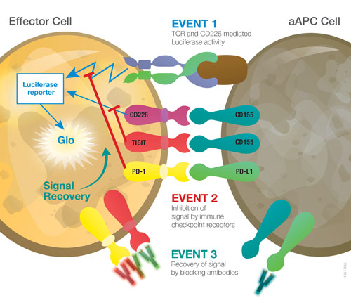
Here, we describe the development of three bioluminescent reporter-based bioassays that provide a quantitative measure of the biological activity of antibodies designed to block PD-1/PD-L1 and TIGIT/CD155, individually and in combination. The assays consist of two cell lines representing T effector cells and antigen-presenting cells (Figure 1). The T effector cells stably express a luciferase reporter that is activated downstream of TCR or CD226 engagement. These cells are then individually engineered to express A) PD-1, B) TIGIT or C) both receptors. The antigen-presenting cells (artificial antigen-presenting cells, or “aAPC”) stably express a T cell activator protein that binds and activates the T effector cell in an antigen-independent manner. The aAPC cells are further engineered to express the ligands for A) PD-L1, B) CD155, or C) both as complementary cell lines to the effector cell lines. When the T effector cell is co-cultured with its corresponding aAPC cell, the PD-1/PD-L1, TIGIT/CD155, or both receptor/ligand interactions inhibit T effector cell activation and luciferase activity. Addition of a relevant anti-PD-1 blocking antibody, anti-TIGIT blocking antibody, or both, releases the inhibitory signal allowing expression of luciferase activity (Figure 1).

Figure 1. Schematic representation of the blockade bioassays for PD-1/PD-L1, TIGIT/CD155, and PD-1+TIGIT. Each bioassay consists of a T effector cell (expressing PD-1, TIGIT, or PD-1+TIGIT) and an aAPC cell (expressing PD-L1, CD155, or PD-L1+CD155), respectively. Co-culture of a T effector cell with its corresponding aAPC cell leads to three events as noted in the image.
Simple & Specific Blockade Bioassays for PD-1/PD-L1 and TIGIT/CD155
The PD-1/PD-L1 and TIGIT/CD155 blockade bioassays use the same simple assay format and demonstrate specificity for the target immune checkpoint receptor/ligand interaction. To demonstrate specificity of the PD-1/PD-L1 blockade bioassay, we first plate aAPC for PD-L1 in cell recovery medium in white, flat-bottom 96-well assay plates. After overnight incubation, we remove the medium and add serial dilutions of anti-PD-1 antibody (nivolumab) to the assay plates, followed by addition of PD-1 effector cells. After six hours of incubation at 37°C, we add Bio-Glo™ Reagent to the assay plates and measure luminescence using a luminescence plate reader (e.g., GloMax® Discover). We used an identical protocol to demonstrate specificity of the TIGIT/CD155 blockade bioassay with an anti-TIGIT antibody.
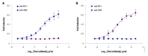
The PD-1/PD-L1 blockade bioassay showed a dose-dependent increase in luciferase activity in response to the anti-PD-1 antibody, nivolumab, but not to an anti-TIGIT antibody (Figure 2A). Conversely, the TIGIT/CD155 blockade bioassay showed a dose-dependent increase in luciferase activity in response to the anti-TIGIT antibody, but not to nivolumab (Figure 2B).
In a subsequent qualification study designed according to ICH guidelines, the PD-1/PD-L1 blockade bioassay showed the appropriate assay precision, accuracy and linearity (data not shown). The simplicity and flexibility of the format of the PD-1/PD-L1 blockade bioassay allows its use in 384- and 1536-well assay plates. As the bioassay is robust, data from any plate-well format are comparable. Together, the data demonstrate the potential use of bioluminescent reporter-based bioassays in immunotherapy drug development, including early drug screening, characterization, potency, and stability testing.

Figure 2. Specificity of the PD-1/PD-L1 and TIGIT/CD155 blockade bioassays. aAPC cells for PD-L1 (A) or CD155 (B) were plated in white, flat-bottom, 96-well assay plates. After overnight incubation at 37°C, the medium was removed and effector cells for PD-1 (A) or TIGIT (B) were added to the assay plates along with increasing concentrations of the respective antibodies, nivolumab (anti-PD-1 antibody) or anti-TIGIT antibody. After six hours, Bio-Glo Reagent was added to the assay plates and luminescence was measured using a luminescence plate reader (e.g., GloMax Discover). Data were analyzed using GraphPad Prism® software.
Synergy of the PD-1+TIGIT Combination Bioassay
The PD-1+TIGIT Combination Bioassay uses the same assay principle as the above blockade bioassays. Here, the T effector cells co-express PD-1 and TIGIT (PD-1+TIGIT effector cells), and aAPC cells co-express PD-L1 and CD155 (PD-L1+CD155 aAPC cells). We engineered PD-1 and TIGIT in the effector cells, and PD-L1 and CD155 in the aAPC cells to control relative expression levels of the receptors, which we confirmed by flow cytometry (data not shown).
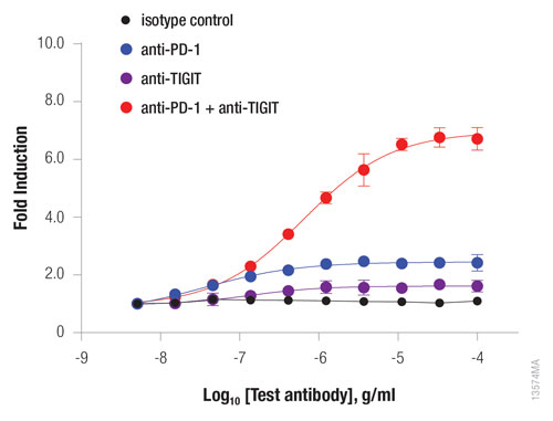
The PD-1+TIGIT combination bioassay showed a twofold, dose-dependent increase in luciferase activity in response to either nivolumab or an anti-TIGIT antibody (Figure 3). Of note, the addition of a 1:1 ratio of anti-PD-1 and anti-TIGIT antibodies resulted in an eightfold increase in luciferase activity. Therefore, the bioluminescent reporter-based PD-1+TIGIT combination bioassay provides a quantitative measure of the synergetic effect of anti-PD-1 and anti-TIGIT immune checkpoint blocking antibodies on T cell activation.

Figure 3. Synergy of anti-PD-1 and anti-TIGIT blocking antibodies measured using the PD-1+TIGIT combination bioassay. PD-1+TIGIT effector cells were incubated with PD-L1+CD155 aAPC cells in the presence of anti-PD-1, anti-TIGIT, or both blocking antibodies. Luciferase activity and data were analyzed as in Figure 2.
Summary
All three bioluminescent reporter-based blockade bioassays, PD-1/PD-L1, TIGIT/CD155, and PD-1+TIGIT, reflect the mechanism of action of antibody drug candidates in development. The bioassays exhibit specificity, simplicity, and robustness with the appropriate assay precision, accuracy, and linearity required for antibody potency and stability testing. Using specific immune checkpoint blocking and immunostimulatory antibodies illustrates the value of reporter-based bioassays for antibody screening and characterization, and each bioassay can be applied during antibody development and manufacture in immunotherapy drug development programs.
FOR RESEARCH USE ONLY.
Zhi-jie Jey Cheng ([email protected]) is sr. research scientist II and group leader, Jamison Grailer is sr. research scientist I, Jim Hartnett is sr. research scientist II, Frank Fan is sr. director, research, and Mei Cong is sr. research manager at Promega.


