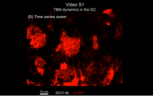
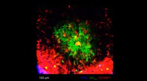
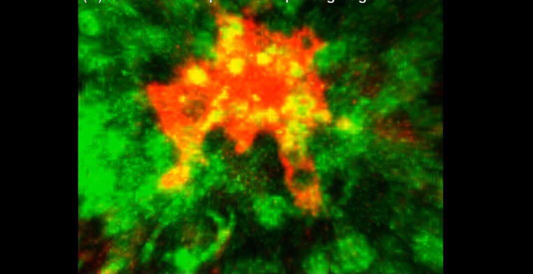
During an antibody response, sites of massive cell death arise in the germinal centers of lymph nodes. One of the macrophage cell types, tingible body macrophages (TBMs), help with apoptotic cell clearance, preventing secondary necrosis and autoimmune activation. Now, for the first time, scientists have tracked the TBM’s life cycle and function, with implications for a deeper understanding of autoimmune disorders.
This work is published in Cell in the paper, “Apoptotic cell fragments locally activate tingible body macrophages in the germinal center.”

During an immune response, a massive number of B cells are made inside the lymph nodes and then tested for their ability to neutralize the infection. B cells that fail the test are destined to die, but on the way out, they can trigger the body to attack itself. The contents of these cells—especially those in the cell’s central nucleus—are inflammatory and can inadvertently activate some B cells to make antibodies against that waste, leading to autoimmunity. Removing this waste is therefore a critical housekeeping function.
“In living organisms, death happens all the time—and if you don’t clean up, the contents of the dead cells can trigger autoimmune diseases,” said Tri Phan, PhD, professor and head of the Intravital Microscopy and Gene Expression (IMAGE) Lab and co-lead of the Precision Immunology Program at Garvan Institute of Medical Research in Australia.
Researchers used intravital imaging techniques to observe how macrophages form within the lymph nodes and how they behave in real time.
First, they found that TBMs “derive from a lymph node-resident, CD169-lineage, CSF1R-blockade-resistant precursor that is prepositioned in the follicle.” They also uncovered that, unlike typical macrophages, “non-migratory TBMs use cytoplasmic processes to chase and capture migrating dead cell fragments using a ‘lazy’ search strategy.”
“We know so very little about tingible body macrophages because it was not possible until now, with next-generation two-photon microscopes, to get inside the microstructures inside the lymph nodes of a living animal and watch the cells in action in real time. That’s why it’s taken 140 years—from when tingible body macrophages were first described in 1885—to get where we are now,” said Phan.

“This research is exciting because it helps us to understand causes of autoimmune conditions like lupus. Understanding why somebody gets the disease in the first place and why it keeps coming back, is an important step towards future treatments for these diseases,” said Wunna Kyaw, a PhD student at Garvan and co-first author of the study.
In systemic lupus, the immune system struggles to control the production of its T cells and B cells. Their overactivity causes inflammation, autoantibodies, and long-term damage throughout the body. This research shows that tingible body macrophages, with their B cell clean-up function, could be responsible for setting the chain of events in motion if they fail.
So far, the study has examined what happens with the macrophages in animal models of a healthy system. The researchers’ next step is to expand the experiment to an autoimmune model, to see if they can rescue the failing system and prevent autoimmunity at its root cause.


