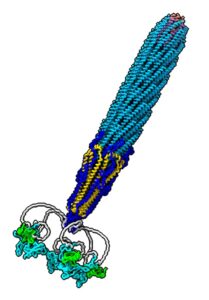
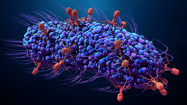
For the first time, researchers at the University of Exeter, in collaboration with Massey University and Nanophage Technologies, have mapped out what a commonly-used form of phage looks like. Their findings may help researchers design better uses of the viruses in biotech.
Their study is published in Nature Communications in an article titled, “Cryo-electron microscopy of the f1 filamentous phage reveals insights into viral infection and assembly.”
“Phages are viruses that infect bacteria and dominate every ecosystem on our planet. As well as impacting microbial ecology, physiology, and evolution, phages are exploited as tools in molecular biology and biotechnology,” the scientists wrote.
“This is particularly true for the Ff (f1, fd, or M13) phages, which represent a widely distributed group of filamentous viruses. Over nearly five decades, Ffs have seen an extraordinary range of applications, yet the complete structure of the phage capsid and consequently the mechanisms of infection and assembly remain largely mysterious. In this work, we use cryo-electron microscopy and a highly efficient system for production of short Ff-derived nanorods to determine a structure of a filamentous virus including the tips. We show that structure combined with mutagenesis can identify phage domains that are important in bacterial attack and for release of new progeny, allowing new models to be proposed for the phage lifecycle.”
“Phages form part of a very exciting and growing area of research, with a range of current and potential applications,” explained Vicki Gold, PhD, a senior lecturer at the University of Exeter. “Yet until now, we’ve not had a complete picture of what filamentous phages look like. We’ve now provided the first view, and understanding this will help us improve applications for phage into the future.”
Because filamentous phages are so long, scientists have previously failed to capture an image of their entirety.

The researchers created smaller versions, which are around 10-fold shorter, and look like straight nanorods rather than entangled spaghetti-like filaments. This mini version was small enough to be imaged in its entirety using high-resolution cryo-electron microscopy.
“Near-atomic resolution structures of filamentous phage tips in conjunction with structure-function analyses is paradigm-shifting for virology,” the scientists wrote. “We reveal how an intertwined network of α-helices forms a highly stable filament, culminating with a pentameric bundle at the leading tip of the infecting phage. This knowledge allows the mechanism of cellular-attack to be rationalized and can now be exploited for mechanistic understanding of the infection and assembly/egress of all filamentous phages.”
In addition, the structures will allow expansion of biotechnological and nanotechnological applications by allowing the precise structure-guided design of novel modification points and 3D display structures.


