October 15, 2010 (Vol. 30, No. 18)
CellASIC Technology Mimics Physiological Tissue Transport Using Continuous Perfusion Systems
Modern biology is driven by technology, explains Philip J. Lee, Ph.D., co-founder, president, and director of research at CellASIC a five-year-old company that develops microfluidic technologies for cell culture.
“While the last 50 years were dominated by gene and protein analysis, the current emphasis is to understand the cell as a system, and the next 50 years will focus on how cells interact to form functional tissues. CellASIC’s motivation is to determine how the need for cell-relevant environments for biologic studies can be met by engineering.”
In 2003 Dr. Lee and CellASIC co-founder Paul Hung, Ph.D., were graduate students in the laboratory of Luke Lee, Ph.D., a professor in the bioengineering department at the University of California, Berkeley. They began to utilize microfabrication methods to improve laboratory cell culture methods. What began as a quest to engineer a “better Petri dish” eventually led to experimenting with a continuous perfusion system that mimicked physiological tissue transport conditions with the ultimate goal of improving live-cell imaging.
“Live-cell imaging is the best way to monitor cells in vitro, because it allows for intracellular resolution and kinetic and spatial detail,” Dr. Philip Lee says. “While cells are dynamic systems that respond to changes in their environment, most studies cannot apply precise time-varying stimuli.”
Borrowing tools and technology from the semiconductor industry, CellASIC’s founders developed the capability to engineer structures on a microscale. They discovered that microfluidic technology enables the delivery of nanoliter volumes to individual units of an array, providing microenvironment control, reduction of sample volumes, inexpensive process automation, and the optical quality to monitor cell conditions in vitro. While microfluidic culture is “fundamentally different from dish-based cell culture,” it adapts to existing biological protocols, according to Dr. Lee.
“Advantages of microfluidic cell culture include creating better microenvironments that serve as models for complex cell behaviors, enabling integration and increased complexity in cell biology, and making live-cell studies easier with novel types of experimentation and more uniform experiment conditions, reduced cost, labor, and reagent use.”
In 2005 CellASIC rented a small laboratory near the university and obtained funding by writing research grants. While the company still gets a large portion of its funding from SBIRs, it is now generating product revenue from its first product, ONIX, launched in 2007. The ONIX microfluidic perfusion platform delivers a high level of control for live-cell imaging experiments. The platform integrates with users’ existing inverted microscope systems to enable dynamic time-lapse experiments. Its flow control system allows for computer-controlled dynamic flow switching for time-lapsed live-cell microscopy.
The ONIX platform’s single-use microfluidic plates work with two independent flow units with identical flow properties to enable simultaneous imaging of two sets of cell/medium combinations. The yeast cell microfluidic plate is optimized for time-lapsed imaging of yeast cells with solution exchange. The microfluidic cell-trapping region holds yeast cells in a uniform focal plane for time-lapse cell microscopy during perfusion flow. The mammalian cell microfluidic plate enables time-lapsed imaging of cultured mammalian cells with solution switching. The microfluidic cell culture region ensures optimal cell health during long-term microscopy studies.
Dr. Lee says the ONIX system is easy to use, with less than 10 minutes required to start collecting data. It is also compatible with existing cell culture workflow. To use ONIX, the researcher pipettes the cell suspension onto a microfluidic plate using capillary-driven cell loading, cultures the cells in a standard incubator via gravity-driven perfusion if desired, pipettes the media and solutions into flow inlet wells, seals them to the ONIX manifold, places the plate on an inverted microscope stage, specifies the solution exposure profile, and performs live-cell imaging.

CellASIC’s newest product is a microfluidic perfusion array that can reportedly maintain liver-specific activity in cultured primary hepatocytes for more than 12 days after plating.
Gel Imaging
CellASIC’s second product is the 3D:M microfluidic cell culture array, which performs perfusion culture in 3-D gel format. There are 24 independent units per 96-well plate, and no external hardware is required. Used in such applications as cancer cell studies, drug screening, and culturing cells in a 3-D environment, the 3D:M enables high-quality gel imaging and uses 100 times less gel. “The result is improved throughput, reduced reagent usage, better data quality, and reduced cost,” Dr. Lee said.
CellASIC’s newest product is a microfluidic perfusion array capable of maintaining liver-specific activity in cultured primary hepatocytes for more than 12 days after plating. The microfluidic hepatocyte array enables improved culture of primary human hepatocytes for in vitro drug evaluation studies of metabolic activity and long-term drug toxicity, according to Dr. Lee. In addition, “the system is a liver-like environment with blood and nutrients perfusing through it to achieve long-term viability and realistic morphology.”
Using microfabrication technology to create a cell environment similar to the liver acinus, the system arranges hepatocytes in parallel, 3-D, plate-like configurations. Each culture region is separated by a fluidic sinusoid that enhances mass transport to the cells, and the exposure solution is continuously perfused to the cells at a rate of 100 microliters per day using a passive gravity method. To allow for operation with standard equipment and automated instruments, the microfluidic hepatocyte plates are formatted to a standard 96-well layout.
“We believe this system will offer a new avenue for more clinically relevant liver-related studies, offering the benefits of improved long-term function, reduction of cell usage, and higher throughput studies,” said Dr. Lee.
In the near term, CellASIC hopes to “educate the resesearch population about using microfluidic technologies for studying cells in vitro and controlling the environment” while finding new applications for CellASIC systems. “As the number of applications increases, microfluidic cell culture will become a more commonplace idea.”

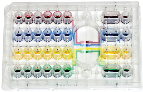
ONIX microfluidic experiment plates were designed for use in live-cell perfusion imaging. They feature crystal clear optics with a 170 µm glass slide, and they can image multiple microchambers in parallel.


