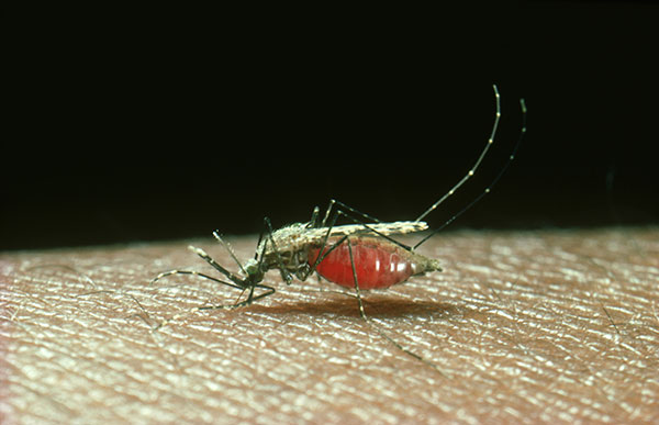
To understand parasitism, one needs to comprehend the often dual nature of many parasitic organisms. For instance, the malaria parasite (Plasmodium) has a two-stage lifecycle where it interacts with drastically different hosts—one always being the female mosquito. The parasites go through distinctly different stages of life in the mosquito hosts, which has been an area of continued study for decades, especially when focused on how the mosquito immune system fights off the malaria parasite.
Now, new evidence from researchers at Iowa State University (ISU) may shine some much-needed light on mosquitos’ immune response mechanisms—potentially laying the groundwork for future research to combat the transmission of malaria. Findings from the new study—published recently in PNAS through an article titled “Chemical depletion of phagocytic immune cells in Anopheles gambiae reveals dual roles of mosquito hemocytes in anti-Plasmodium immunity”—show how the mosquito immune system combats malaria parasites at multiple stages of development.
“Mosquitoes are generally pretty good at killing off the parasite,” remarked senior study investigator Ryan Smith, PhD, assistant professor of entomology at ISU. “We wanted to figure out the mechanisms and pathways that make that happen.”
Roughly 219 million cases of malaria, a disease transmitted to humans by the bite of infected mosquitoes, occurred worldwide in 2017, according to the Centers for Disease Control and Prevention. Most cases are concentrated in tropical and subtropical climates such as sub-Saharan Africa and South Asia. The disease resulted in 435,000 deaths in 2017, according to the CDC.
Mosquitoes are required to transmit malaria as they acquire the parasites by biting an infected person and then transmit the disease later after the parasite has completed its development within the mosquito. This new study focused on how the mosquito immune system responds to the parasite.
The researchers treated mosquitoes with a chemical that depleted their immune cells, which are needed to defend the mosquito against pathogens. The experiments showed that malaria parasites survived at greater rates in mosquitoes when the immune cells were depleted. Moreover, the research also illuminated how these immune cells promoted different “waves” of the mosquito immune response targeting distinct stages of malaria parasites in the mosquito host.
“The lack of genetic tools has limited hemocyte study despite their importance in mosquito anti-Plasmodium immunity, the authors wrote. “To address these limitations, we employ the use of a chemical-based treatment to deplete phagocytic immune cells in Anopheles gambiae, demonstrating the role of phagocytes in complement recognition and prophenoloxidase production that limit the ookinete and oocyst stages of malaria parasite development, respectively. Through these experiments, we also define specific subtypes of phagocytic immune cells in An. gambiae, providing insights beyond the morphological characteristics that traditionally define mosquito hemocyte populations.”
Smith, who also leads the ISU Medical Entomology Laboratory, said the findings increase the understanding of a complement-like pathway that is involved in the initial recognition and killing of parasites, similar to that found in mammals. The work also implicates phenoloxidases, an insect-specific immune response, in causing a secondary immune response directed at later stages of the malaria parasite, he said.
Understanding these immune responses could lead to opportunities to eliminate malaria parasites in the mosquito, thus reducing the transmission of malaria. For instance, Smith said scientists could use genetic approaches to make mosquitoes resistant to malaria parasites. Introducing mosquitoes with enhanced immunity in endemic areas of malaria could significantly reduce human malaria cases.
“There are more steps required to validate that kind of approach, but we think this study lays a foundation for those future experiments,” Smith concluded.


