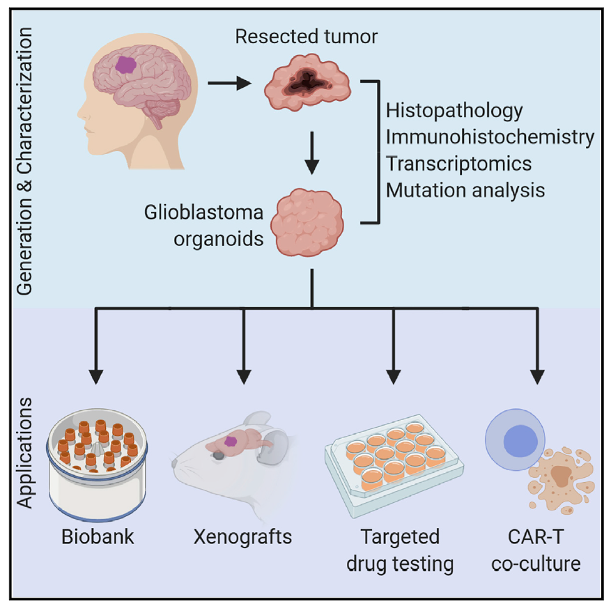
Organoids are rapidly becoming an integral part of the drug discovery process and now investigators at the University of Pennsylvania School of Medicine believe that lab-grown brain organoids developed from a patient’s own glioblastoma, the most aggressive and common form of brain cancer, may hold the answers on how to best treat it. The researcher’s findings—published recently in Cell through an article entitled “A Patient-Derived Glioblastoma Organoid Model and Biobank Recapitulates Inter- and Intra-tumoral Heterogeneity”—shows how glioblastoma organoids could serve as effective models to rapidly test personalized treatment strategies.
Glioblastoma multiforme (GBM) remains the most difficult of all brain cancers to study and treat, largely because of tumor heterogeneity. Treatment approaches, like surgery, radiation, and chemotherapy, along with newer personalized cellular therapies, have proven to slow tumor growth and keep patients disease-free for some periods of time—however, a cure remains elusive.
“While we’ve made important strides in glioblastoma research, preclinical and clinical challenges persist, keeping us from getting closer to more effective treatments,” explained senior study investigator Hongjun Song, PhD, professor of neuroscience in the Perelman School of Medicine at the University of Pennsylvania. “One hurdle is the ability to recapitulate the tumor to not only better understand its complex characteristics, but also to determine what therapies post-surgery can fight it in a timelier manner.”

Amazingly, what makes organoids so attractive in GBM, is timing and the ability to maintain cell type and genetic heterogeneity. While existing in vitro models have added to researchers’ understanding of the biological mechanisms underlying the cancer, they have limitations. Unlike other models, which need more time to exhibit gene expression and other histological features that more closely represent the tumor, brain tumor organoids developed by the research group grow into use much more rapidly. That’s important because current treatment regimens are typically initiated one month following surgery, so having a road map sooner is more advantageous.
In the current study, the researchers removed fresh tumor specimens from 52 patients to “grow” corresponding tumor organoids in the lab. The overall success rate for generating glioblastoma organoids (GBOs) was 91.4%, with 66.7% of tumors expressing the IDH1 mutation, and 75% for recurrent tumors, within two weeks. These tumor glioblastoma organoids can also be biobanked and recovered later for analyses.
Genetic, histological, and molecular analyses were also performed in 12 patients to establish that these new GBOs had largely retained features from the primary tumor in the patient. Eight GBO samples were then successfully transplanted into adult mouse brains, which displayed rapid and aggressive infiltration of cancer cells and maintained key mutation expression up to three months later. Importantly, a major hallmark of GBM—the infiltration of tumor cells into the surrounding brain tissue—was observed in the mouse models.
To mimic post-surgery treatments, the researchers subjected GBOs to standard-of-care and targeted therapies, including drugs from clinical trials and chimeric antigen receptor T (CAR-T) cell immunotherapy. For each treatment, researchers showed that the organoid responses are different, and effectiveness is correlated to their genetic mutations in patient tumors. This model opens the possibility for future clinical trials for personized treatment based on individual patient tumor responses to various drugs.
Notably, the researchers observed a benefit in the organoids treated with CAR-T therapies, which have been used in ongoing clinical trials to target the EGFRvIII mutation, a driver of the disease. In six GBOs, the researchers showed the specific effect to patient GBOs with the EGFRvIII mutation with an expansion of CAR-T cells and reduction in EGFRvIII expressing cells.
“These results highlight the potential for testing and treating glioblastomas with a personalized approach. The ultimate goal is to work towards a future where we can study a patient’s organoid and test which CAR-T cell is going to be the best against their tumor, in real-time,” concluded co-senior study investigator Donald O’Rourke, MD, professor in neurosurgery, and director of the GBM Translational Center of Excellence at Penn’s Abramson Cancer Center. “A shorter-term goal, given the heterogeneity of glioblastomas, is that in vitro testing of various therapeutic options may also help refine patient enrollment in clinical trials, by more accurately defining mutations and selecting the appropriate, available targeted therapies for each.


