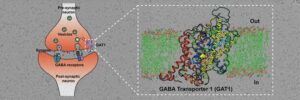
Researchers at the Indian Institute of Science and collaborators used cryo-EM to decode the molecular architecture of a transporter protein controlling the movement of gamma-aminobutyric acid (GABA). GABA is an inhibitory neurotransmitter that balances out the excitatory inputs from glutamate, another neurotransmitter.
A balance between excitation and inhibition is vital for the neural circuitry to maintain normal structure and function. Imbalances in excitatory or inhibitory inputs can result in disorders like seizures, anxiety, and schizophrenia.
The scientists published their study, “Cryo-EM structure of GABA transporter 1 reveals substrate recognition and transport mechanism,” in Nature Structural & Molecular Biology.
“The inhibitory neurotransmitter γ-aminobutyric acid (GABA) is cleared from the synaptic cleft by the sodium- and chloride-coupled GABA transporter GAT1. Inhibition of GAT1 prolongs the GABAergic signaling at the synapse and is a strategy to treat certain forms of epilepsy. In this study, we present the cryo-electron microscopy structure of Rattus norvegicus GABA transporter 1 (rGAT1) at a resolution of 3.1 Å,” the investigators wrote.
Epitope transfer
“The structure elucidation was facilitated by epitope transfer of a fragment-antigen binding (Fab) interaction site from the Drosophila dopamine transporter (dDAT) to rGAT1. The structure reveals rGAT1 in a cytosol-facing conformation, with a linear density in the primary binding site that accommodates a molecule of GABA, a displaced ion density proximal to Na site 1 and a bound chloride ion. A unique insertion in TM10 aids the formation of a compact, closed extracellular gate.
“Besides yielding mechanistic insights into ion and substrate recognition, our study will enable the rational design of specific antiepileptics.”

GABA transporters (GATs) are the primary molecules involved in this step—they employ sodium and chloride ions to move excess GABA back into the neurons. GATs are therefore vital molecules that orchestrate GABA signaling and function. They are, therefore, an important target for the treatment of conditions like seizures.
The team, led by Aravind Penmatsa, PhD, associate professor in the molecular biophysics unit, purified GAT and used a novel approach to create an antibody site on this molecule. Antibodies help increase the mass of proteins and facilitate improved imaging through cryo-EM. The researchers were able to observe that the GAT structure was facing the cytosol (the inside of the cell) and was bound to a GABA molecule, sodium, and chloride ions. This binding is one of many key steps in the GABA transport cycle; deciphering it can provide vital insights into the mechanisms of GABA recognition and release into neurons.
The availability of high-resolution GAT structures is crucial for developing specific blockers of GABA uptake for treatment of epilepsies. It would also aid in studying how drugs prescribed to block GABA uptake interact with GATs.


