The discovery of COVID-19 treatments is urgently needed. As part of that process, so too are appropriate animal models to test them. However, which animal(s) can be the most useful in this scientific endeavor remains a question.
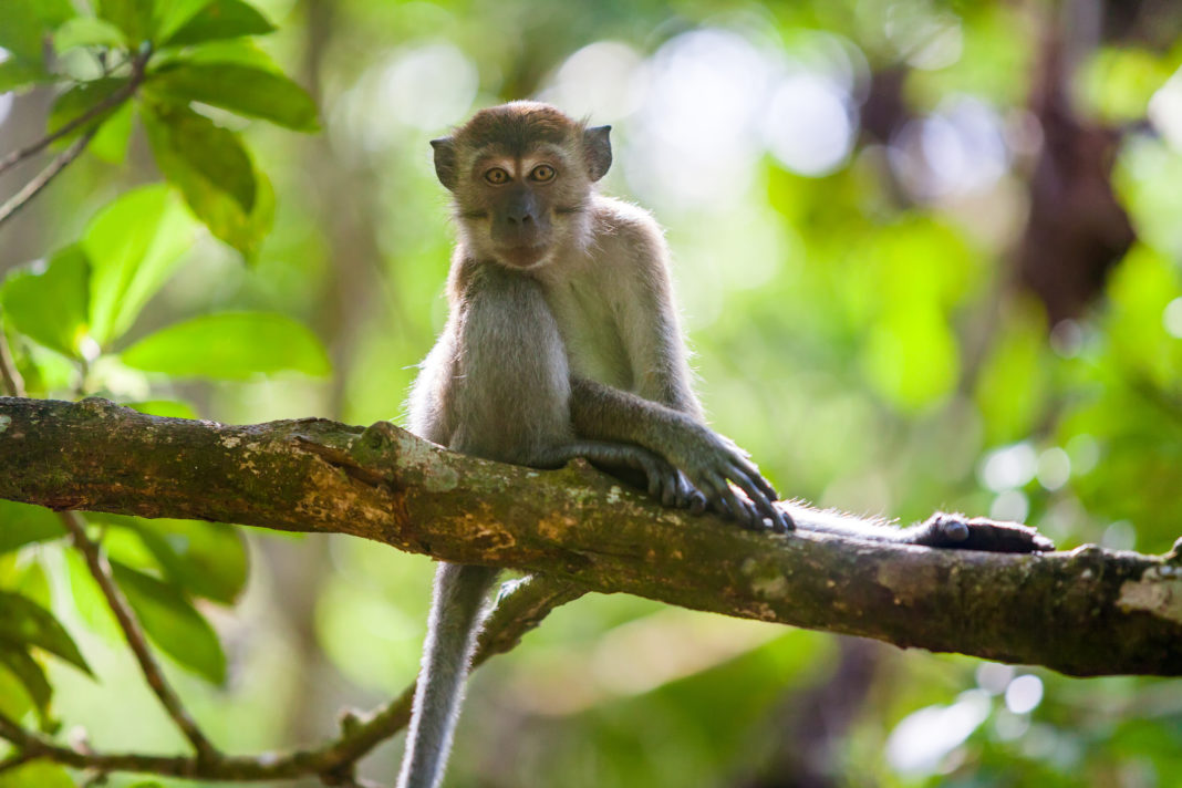
A group from the Netherlands sought to address this question. They infected young and aged cynomolgus macaques with a clinical isolate of SARS-CoV-2. The pathogenesis of SARS-CoV infection had been previously studied in this nonhuman primate model. Not only did they characterize the infection of SARS-CoV-2, they also compared that infection with that of MERS-CoV, and results with historical reports of infections by SARS-CoV.
The work was recently published in Science in the paper titled, “Comparative pathogenesis of COVID-19, MERS, and SARS in a nonhuman primate model.”
The researchers inoculated “two groups of four cynomolgus macaques (both young adult (4–5 years of age); and old adult (15–20 years of age) by a combined intratracheal (IT) and intranasal (IN) route with a SARS-CoV-2 strain from a German traveler returning from China.”
SARS-CoV-2 led to mild infection in cynomolgus macaques with little to no symptoms, the authors reported, even though animals infected were shedding the virus. Despite the absence of clinical signs, the animals shed the virus, as measured by RT-qPCR and virus culture of nasal, throat, and rectal swabs. The authors noted that, in nasal swabs, “detection of SARS-CoV-2 RNA peaked by day 2 post-infection (pi) in young animals, by day 4 pi in aged animals, was detected up to at least day 8 pi in two out of four animals and up to day 21 pi in one out of four animals.”
This has interesting similarities to how asymptomatic humans shed the SARS-CoV-2 virus, such as the human presymptomatic and asymptomatic cases that have also shed virus.
None of the infected animals exhibited overt clinical signs or significant weight loss, even though all of the animals produced SARS-CoV-2 specific antibodies. Increased age did not affect disease outcome, but there was prolonged viral shedding in the upper respiratory tract of aged animals—an observation that has also been made in both SARS-CoV-2 and SARS-CoV human patients.
Interestingly, SARS-CoV-2 shedding in the asymptomatic model peaked early in the course of infection, similar to what is seen in symptomatic patients. SARS-CoV-2 was primarily detected in tissues of the respiratory tract, however, viral RNA was also detectable in other tissues such as intestines.
Comparing viruses
To better understand key characteristics in the pathogenesis of SARS-CoV-2, the team compared the cynomolgus macaques infected with SARS-CoV-2 to those infected with MERS-CoV and compared the pathology and virology with historical reports of SARS-CoV infections.
Like influenza, the animals infected with SARS-CoV-2 shed the virus from the respiratory tract very early during infection as compared to SARS-CoV, which may be part of the explanation of the explosive global spread of COVID-19 and why case detection and isolation may not be as effective as it was for controlling SARS-CoV.
Whereas MERS-CoV primarily infects type II pneumocytes in cynomolgus macaques, both SARS-CoV and SARS-CoV-2 also infect type I pneumocytes. Injury to type I pneumocytes can result in pulmonary edema, and formation of hyaline membranes which, the authors noted, “may explain why hyaline membrane formation is a hallmark for SARS and COVID-19 but not frequently reported for MERS.”
After comparing how the coronavirus infections develop in cynomolgus macaques, the researchers reported that SARS-CoV-2 gives the animals a mild COVID-19-like disease and suggest that these animals are a promising model for testing COVID-19 therapeutics.
“This study provides a novel infection model,” the authors wrote, “which will be critical in the evaluation and licensure of preventive and therapeutic strategies against SARS-CoV-2 infection for use in humans.”


