Seeing is believing! Yet, in science, it’s not always practical or even possible to visualize events that occur in the natural world. Take, for instance, the human brain—an organ heavily shielded for its protection and often shrouded in mystery. While neuroscience has exponentially improved in the past few decades, visualization of various neuronal events has been exceedingly difficult. However now, investigators at The European Molecular Biology Laboratory (EMBL) have just captured, for the first time, microglia cells nibbling on brain synapses. Findings from the new study—published today in Nature Communications, in an article entitled “Microglia Remodel Synapses by Presynaptic Trogocytosis and Spine Head Filopodia Induction”—show that the special glial cells help synapses grow and rearrange, demonstrating the essential role of microglia in brain development.
“Our findings suggest that microglia are nibbling synapses as a way to make them stronger, rather than weaker,” explained senior study investigator Cornelius Gross, Ph.D., the deputy head of outstation and senior scientist at EMBL.
Around one in ten cells in your brain are microglia. Related to macrophages, they act as the first and main contact in the central nervous system's active immune defense. They also guide healthy brain development. Researchers have proposed that microglia pluck off and eat synapses—connections between brain cells—as an essential step in the pruning of connections during early circuit refinement. But, until now, no one had seen them do it.
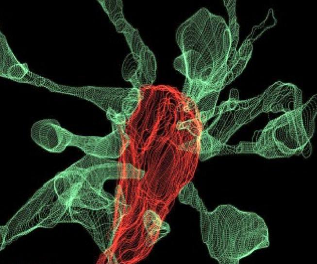
The EMBL researchers saw that roughly half of the time that microglia contact a synapse, the synapse head sends out thin projections or “filopodia” to greet them. In one particularly dramatic case—as seen in the image above—fifteen synapse heads extended filopodia toward a single microglia cell.
“As we were trying to see how microglia eliminate synapses, we realized that microglia actually induce their growth most of the time,” noted lead study investigator Laetitia Weinhard, Ph.D., a postdoctoral research scientist at EMBL.
Amazingly, it turns out that microglia might underlie the formation of double synapses, where the terminal end of a neuron releases neurotransmitters onto two neighboring partners instead of one. This process can support effective connectivity between neurons.
“This shows that microglia are broadly involved in structural plasticity and might induce the rearrangement of synapses, a mechanism underlying learning and memory,” Dr. Weinhard remarked.
Since this was the first attempt to visualize this process in the brain, the current study entails five years of technological development. The research team tried three different state-of-the-art imaging systems before succeeding.
“Here we used light sheet fluorescence microscopy to follow microglia–synapse interactions in developing organotypic hippocampal cultures, complemented by a 3D ultrastructural characterization using correlative light and electron microscopy (CLEM),” the authors wrote. “Our findings define a set of dynamic microglia–synapse interactions, including the selective partial phagocytosis, or trogocytosis (trogo-: nibble), of presynaptic structures and the induction of postsynaptic spine head filopodia by microglia. These findings allow us to propose a mechanism for the facilitatory role of microglia in synaptic circuit remodeling and maturation.”
By CLEM and light sheet fluorescence microscopy—a technique developed at EMBL—they researchers were able to make the first movie of microglia eating synapses.
“This is what neuroscientists have fantasized about for years, but nobody had ever seen before,” Dr. Gross concluded. “These findings allow us to propose a mechanism for the role of microglia in the remodeling and evolution of brain circuits during development.” In the future, he plans to investigate the role of microglia in brain development during adolescence and the possible link to the onset of schizophrenia and depression.


