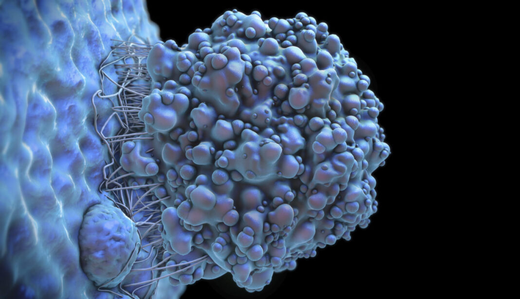
Leptomeningeal metastases (LM) occur when cancer spreads to the membranes lining the brain and spinal cord. The leptomeninges are the membranes lining the brain and spinal cord. Cancer cells infiltrate the relatively isolated spaces of the central nervous system filled with cerebrospinal fluid (CSF) and metastasize to the leptomeningeal membranes. The CSF presents a hostile environment to outsiders, but cancer cells still find a way to survive and thrive in the environment. Now researchers from Memorial Sloan Kettering Cancer Center and Columbia University College of Physicians and Surgeons report that the cancer cells that cause LM take over crucial iron micronutrients from native macrophages.
Their study, “Cancer cells deploy lipocalin-2 to collect limiting iron in leptomeningeal metastasis,” is published in Science and led by Adrienne A. Boire, MD, PhD, neuro-oncologist and neurologist at Memorial Sloan Kettering Cancer Center.
The cerebrospinal fluid cushions the brain within the skull and serves as a shock absorber for the central nervous system. It also circulates nutrients and chemicals filtered from the blood and removes waste products from the brain.
“To investigate the mechanism by which cancer cells in these LM overcome these constraints, we subjected CSF from five patients with LM to single-cell RNA sequencing,” the researchers wrote.
The researchers discovered that the cancer cells increase their activity of a gene called Lipocalin-2. Lipocalin-2 (LCN2) is a protein that in humans is encoded by the LCN2 gene. LCN2 induces chemokine production in the CNS in response to inflammatory challenges, and actively participates in the innate immune response, cellular influx of iron, and regulation of neuroinflammation and neurodegeneration. In the CSF, the cancer cells boost their Lipocalin-2 protein levels to outcompete the immune cells for iron in the environment.
“We found that cancer cells, but not macrophages, within the CSF express the iron-binding protein LCN2 and its receptor SCL22A17. These macrophages generate inflammatory cytokines that induce cancer cell LCN2 expression but do not generate LCN2 themselves. In mouse models of LM, cancer cell growth is supported by the LCN2/SLC22A17 system and is inhibited by iron chelation therapy. Thus, cancer cells appear to survive in the CSF by outcompeting macrophages for iron,” noted the researchers.
“It’s nefarious, the way the cancer cells exploit Lipocalin-2 to gain an advantage over the immune cells,” Boire stated.
Because iron is limiting in the CSF, the researchers believed that iron chelation might impair cancer cell growth in the CSF and tested their hypothesis in mouse models. They delivered the chelators directly into the spinal fluid, and found it slowed the growth of the cancer cells.
The team of researchers is looking to bring this therapy to a clinical trial. The work features the remarkable plasticity of tumor cells and reveals potential new avenues for treating these particularly intractable advanced cancer complications. Their findings hold promise as current treatments for leptomeningeal metastasis are not effective at destroying the cancer cells.
“The evolutionary dynamics that we have discovered between malignant and nonmalignant cells within the leptomeninges reveal both the robust nature of cancer’s transcriptional plasticity and microenvironmental vulnerabilities ripe for therapeutic exploitation,” concluded the researchers.


