Researchers at the University of Cambridge have discovered a specific brain region that underlies “goal-directed behavior”—that is, when we consciously do something with a particular goal in mind, say, going to the shops to buy food. The study found that marmoset monkeys could no longer make an association between their behavior and a particular outcome when a region of the brain called the anterior cingulate cortex was temporarily switched off. When this area of the brain was silenced, the animals didn’t change their behavior, even when that behavior stopped having the expected outcome.
The scientists suggest that their findings could represent a step towards the development of targeted treatments for human disorders that manifest with compulsive behavior, such as obsessive compulsive disorder (OCD) and eating disorders, and which are thought to involve impaired function in this brain region. “We have identified the very specific region of the brain involved in goal-directed behavior, said Lisa Duan, PhD, at the University of Cambridge Department of Psychology. “When we temporarily turned this off, behavior became more habitual – like when we go onto autopilot.” Added Trevor Robbins, PhD, at the University of Cambridge Department of Psychology, “this is a first step towards identifying suitable molecular targets for future drug treatments, or other forms of therapy, for devastating mental health disorders such as OCD and addiction.”
Duan is first author, and Robbins is co-senior author, of the team’s published paper in Neuron, which is titled, “Controlling one’s world: identification of sub-regions of primate PFC underlying goal-directed behaviour.”
In our everyday lives we continually make decisions based on our goals, or go into “autopilot” to get through the day, the authors explained. “Normal behaviors can either be goal-directed, when performing an action to obtain a specific goal, or habitual, when a stimulus can trigger a well-learned response regardless of its consequences.” The goal-directed system is needed so that we can adapt and remain flexible to our changing environments and goals, while the “habit” system reduces our cognitive load, the team continued. Problems in the coordination of and competition between the goal-directed and habitual systems can be seen in neuropsychiatric disorders such as OCD and addiction. “Thus, understanding the neurobiological basis of goal-directed behaviors will provide insight into the etiology of such disorders.”
To make optimal choices we need to predict, or to believe, that the action (A) will cause the desired outcome (O), the team noted. So whether or not an outcome is contingent upon an action will depend on the probability of that outcome occurring after the action, but also on the probability of the outcome occurring even if the action isn’t taken.
Previous evidence from patients suffering brain damage, and from brain imaging in healthy volunteers, has shown that part of the brain called the prefrontal cortex (PFC) is involved in goal-directed behavior. However, the prefrontal cortex is a complex structure with many regions, and it has not previously been possible to identify the specific part responsible for goal-directed behaviour from human studies alone.
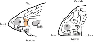
For their reported studies, the University of Cambridge team examined the effects of pharmacologically, and reversibly switching off—or over-activating—different parts of the brain in marmosets. These regions were: areas 24 (perigenual anterior cingulate cortex), 32 (medial PFC), 11 (anterior orbitofrontal cortex, OFC), 14 (rostral ventromedial PFC/medial OFC) and 14–25 (caudal ventromedial PFC), and the anteromedial caudate.

Marmosets were used because their brains share important similarities with human brains, and it is possible to manipulate specific regions of their brains to understand causal effects. “The structural organization of the marmoset PFC bears a greater resemblance to that of humans than to rodents,” the investigators noted, “and hence provides an important bridge between rodent and human studies.”
The marmosets were first taught a goal-directed behavior. By tapping a colored cross when it appeared on a touchscreen, they were rewarded with their favorite juice to drink. But this connection between action and reward was randomly uncoupled, so that they sometimes received the juice without having to respond to the image. The animals quickly detected this change and stopped responding to the image, because they saw they could get juice without doing anything.
Using drugs, the researchers then temporarily inactivated the anterior cingulate cortex, including its connections with another brain region called the caudate nucleus. On repeating the experiment, they found when the connection between tapping the cross and receiving juice was randomly uncoupled, the marmosets did not change their behavior but instead still kept tapping the cross when it appeared.
Such habitual responding to the colored cross was not observed when several other neighboring regions of the brain’s prefrontal cortex—which is known to be important for other aspects of decision-making—were switched off. This indicated the specificity of the anterior cingulate region for goal-directed behavior. “Perigenual ACC (area 24) is the only PFC sub-region in this study that disrupted animals’ sensitivity to the current A-O contingencies when either inactivated or over-activated,” the scientists noted.” Interestingly, a similar effect has been observed in computer-based tests on patients with OCD or addiction: when the relationship between an action and an outcome is uncoupled the patients continue to respond as though the connection is still there.
The results reported by Duan and colleagues may provide new insights into how healthy people behave in a goal-directed way. “We think this is the first study to have established the specific brain circuitry that controls goal-directed behavior in primates, whose brains are very similar to human brains,” said co-senior study author Angela Roberts, PhD, in the University of Cambridge Department of Physiology, Development and Neuroscience. As the investigators further noted, “This study provides the first causal evidence in primates that area 24 (perigenual ACC) is necessary for detecting and acting on changes in instrumental action-outcome (A-O) contingencies and hence the capacity for understanding whether one’s behavior exerts control over the environment.”
The latest study findings are important because the compulsive behaviors in OCD and addiction are thought to result from impairments in the goal-directed system in the brain. In these conditions, worrying, obsessions or compulsive behavior such as drug seeking may reflect an alternative, habit-based system at work in the brain, in which behaviors are not correctly linked with their outcomes. “These findings demonstrate distinct roles of PFC sub-regions in goal-directed behavior and illuminate the candidate neurobehavioral substrates of psychiatric disorders including obsessive-compulsive disorder,” the team wrote.
“Our findings have implications for understanding the neural control of goal-directed behavior and for certain psychiatric disorders, including OCD and schizophrenia.” Studies have previously linked schizophrenia with a loss of GABA-ergic neurons in the anterior cingulate cortex, they pointed out. “… this might be associated with the impairments in goal-directed behavior seen in people with schizophrenia, which may underlie the ‘negative’ symptoms of schizophrenia.”


