Mindy I. Davis Ph.D. National Institute of Health
PLK1 level is elevated in glioblastoma multiforme, and its inhibition restricts the growth of brain cancer cells.
ASSAY & Drug Development Technologies offers a unique combination of original research and reports on the techniques and tools being used in cutting-edge drug development. The journal includes a “Literature Search and Review” column that identifies published papers of note and discusses their importance. GEN presents one article that was analyzed in the “Literature Search and Review” column, a paper published in Stem Cells titled “Polo-like kinase 1 (PLK1) inhibition kills glioblastoma multiforme brain tumour cells in part through loss of SOX2 and delays tumour progression in mice.” Authors of the paper are Lee C, Fotovati A, Triscott J, Chen J, Venugopal C, Singhal A, Dunham C, Kerr JM, Verreault M, Yip S, Wakimoto H, Jones C, Jayanthan A, Narendran A, Singh SK, and Dunn SE.
Abstract from Stem Cells
Glioblastoma multiforme (GBM) ranks amongst the deadliest types of cancer and given this, new therapies are urgently needed. To identify molecular targets, we queried a microarray profiling 467 human GBMs and discovered that polo-like kinase 1 (PLK1) was highly expressed in these tumours and that it clustered with the proliferative subtype. Patients with PLK1-high tumours were more likely to die from their disease suggesting that current therapies are inactive against such tumours. This prompted us to examine its expression in brain tumour initiating cells (BTICs) given their association with treatment failure. BTICs isolated from patients expressed 110–470 times more PLK1 than normal human astrocytes. Moreover, BTICs rely on PLK1 for survival because the PLK1 inhibitor BI2536 inhibited their growth in tumoursphere cultures. PLK1 inhibition suppressed growth, caused G2/M arrest, induced apoptosis and reduced the expression of SOX2, a marker of neural stem cells, in SF188 cells. Consistent with SOX2 inhibition, the loss of PLK1 activity caused the cells to differentiate based on elevated levels of GFAP and changes in cellular morphology.
We then knocked-down SOX2 with siRNA and showed that it too inhibited cell growth and induced cell death. Likewise, in U251 cells, PLK1 inhibition suppressed cell growth, down-regulated SOX2 and induced cell death. Furthermore, BI2536 delayed tumour growth of U251 cells in an orthotopic brain tumour model, demonstrating that the drug is active against GBM. In conclusion, PLK1 level is elevated in GBM and its inhibition restricts the growth of brain cancer cells.
Commentary
The concept of cancer stem cells and the need to target these cells for an effective treatment of cancer has emerged over the past several years. Cancer stem cells are tumor-initiating cells that are in an undifferentiated state and are capable of self-renewal. The authors identify PLK1 (polo-like kinase 1) as a target for the treatment of glioblastoma multiforme based on both siRNA and small molecule inhibition. Glioblastoma is a brain cancer that despite much research still carries a bad prognosis. This is particularly true for the proliferative subtype of the tumors.
Brain tumor initiating cells (BTICs) from patients had several hundred-fold more PLK1 as compared to the levels in normal astrocytes. The presence of PLK1 also correlated with disease severity. BTICs have been recalcitrant to chemotherapy and radiation due to their ability to limit apoptosis, repair DNA damage, and efflux drugs. Treatments that leave the BTICs behind may allow the tumor to relapse.
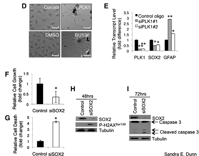
Interestingly, the ATP-competitive PLK1 inhibitor BI2536 led to the differentiation of pediatric SF188 cells (see Figures A–I). This was accompanied by a reduction in the expression of the neural stem cell marker SOX2, which is important for self-renewal of stem cells.
Figures A–C. PLK1 inhibition down-regulates the expression of SOX2, which is required for the growth and survival of GBM cells. (A) SF188 cells (1 × 10⁴ cells per well in 6-well plates) were plated in neurobasal growth medium containing 5 or 10 nM BI2536 for 6 days. The total number of tumourspheres (>50 µm) in each well was counted and photomicrographs of the spheres were taken (scale bar = 500 µm). The treatments were performed in duplicates on three separate occasions. (B) The transcript and protein expression of neural stem cell markers SOX2, musashi, and Bmi1 were measured by RT-PCR and immunoblotting 36 and 48 hours, respectively, after PLK1 siRNA treatment in SF188 cells. (C) The transcript and protein expression of SOX2, musashi, and Bmi1 were measured by RT-PCR and immunoblotting 36 and 48 hours, respectively, after BI2536 treatment in SF188 cells. [Sandra E. Dunn]

Figures A–C
Figures D–I. (D) SF188 cells were treated with 5 nM of PLK1 siRNA or BI2536 for 6 days and photomicrographs were taken on the cells that remained after the treatment. Representative photomicrographs of the cells that underwent dramatic cellular morphological alterations are shown. Scale bar = 280 μm. (E) Total RNA from the cells treated with 5 nM PLK1 siRNA #1 and #2 for 36 hours was extracted and subjected to RT-PCR to quantify the transcripts of PLK1, SOX2, and GFAP. (F) SF188 cells were treated with 100 nM SOX2 siRNA for 72 hrs. The cells were stained with Hoechst and quantified. The number of viable cells was enumerated and the relative fold difference in cell growth is shown in the bar graph. (G) The number of nonviable cells after 100 nM, 72-hour siSOX2 treatment was enumerated based on enhanced Hoechst staining due to chromatin condensation and the relative fold difference in cell death is shown in the bar chart. (H) Proteins extracted from the cells treated with 100 nM control or SOX2 siRNA for 48 hours were subjected to immunoblotting to examine the phosphorylation of H2AX at Ser139, which is a marker of early apoptosis. (I) SF188 cells were treated with 100 nM control or SOX2 siRNA for 72 hours and immunoblotting was performed on the total protein lysates. Increased caspase 3 cleavage was observed in the siSOX2-treated cells compared to the control cells. [Sandra E. Dunn]

Figures D–I
Cell growth was arrested at G2/M and apoptosis was induced. These phenomena were also observed in adult U251 cells and with PLK1 knockdown rather than BI2536 treatment. At low doses (5–10 nM), this drug also blocked the formation of three-dimensional tumorspheres. The ability to dose at low levels would be expected to favor the ability to obtain a therapeutic window. Mice injected intracranially with U251 cells were allowed to develop tumors and were subsequently treated with BI2536 once a week for 4 weeks. The conclusion of the study was that the drug was well tolerated at 50 mg/kg and that there was a significant delay in tumor progression with a corresponding increase in survival.
BI2536 is currently in phase I and II clinical trials for other solid tumors. BI2536 is very selective but not as selective as the exquisitely selective imatinib (S[300 nM] = 0.0337 and 0.0233, respectively) (Davis et al., Nat Biotechnol 2011;29:1046–1051). The ability of BI2536 to address some of the stemness characteristics of BTICs may render these cells more susceptible to chemotherapy and radiation. In a separate study, pretreatment with BI2536 was shown to sensitize the medulloblastoma cell line Daoy, which overexpresses PLK1, to ionizing radiation in addition to reducing colony formation and cell growth, inducing apoptosis and decreasing levels of SOX2 (Harris et al., BMC Cancer 2012;12:80).
The potential to use a combination therapy of traditional chemotherapeutics and/or radiation with BI2536 for the treatment of glioblastoma multiforme and medulloblastoma looks promising.
Mindy I. Davis, Ph.D., works at the NIH.


