One of the driving questions for biologists throughout history has been, “How does the body work?” It is a question that has been answered, albeit at different scales and levels of precision, repeatedly over the course of centuries, from William Harvey’s investigations into the circulatory system to the sequencing of the human genome.
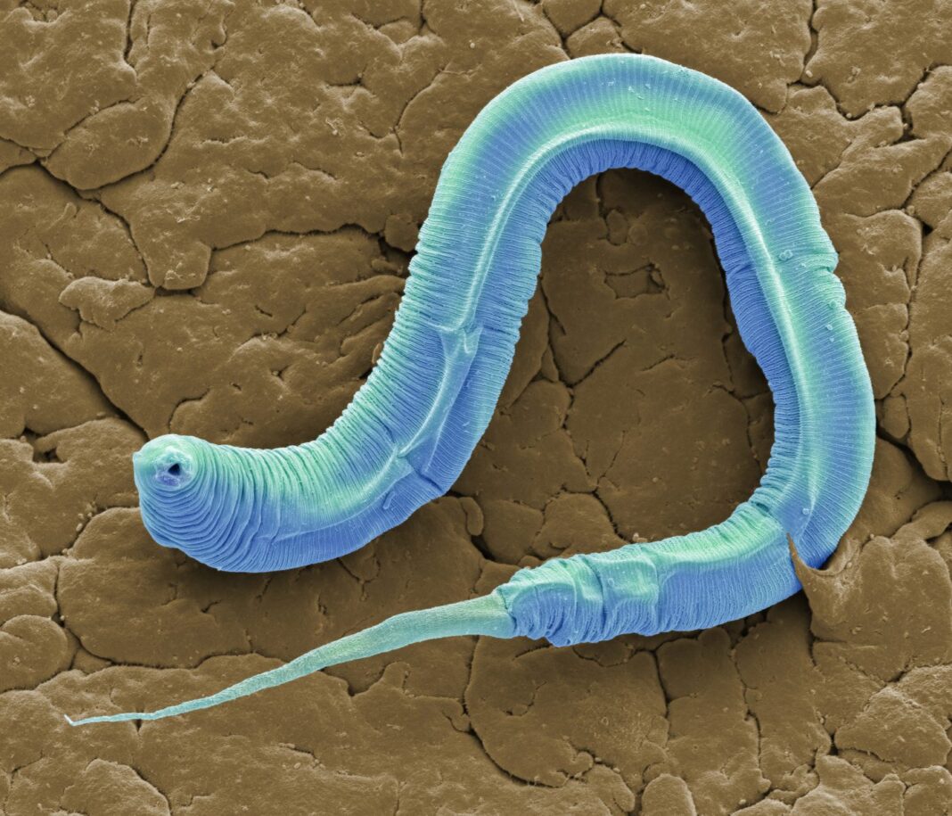
A team of neuroscientists at Albert Einstein College of Medicine have now answered it once again, in spectacular detail. In a recent paper in Nature, they describe the first complete neural wiring diagram, or connectome, for an organism, the nematode roundworm Caenorhabditis elegans. By conceptually simplifying the cells of the worm into a collection of nodes—corresponding to each muscle, organ, or neuron—they were able to chart every synaptic connection between these nodes in both the male and hermaphrodite sexes.
The team, led by Scott Emmons, PhD, professor of genetics, used digitized electron micrographs of C. elegans to reconstruct all 579 nodes of the male worm, including 385 neurons, 155 muscles, and 39 organs, and all 460 nodes of the hermaphrodite worm, with 302 neurons, 132 muscles, and 26 organs. “We have the whole animal, down to every muscle and the connections between the muscles,” he says. “That’s why we use the term whole animal — it covers the whole animal. And then by adding the male to it, of course, now we have the two adult sexes.”
It’s an achievement with far wider impact than just the worm field, however. Deciphering the connections in the worm brain could provide the basis for better understanding the neuroscience and neuropathology of more complex organisms, according to Greg Farber, PhD, director of the Office of Technology Development and Coordination and the National Institute of Mental Health, which supports NIH programs such as the Brain Initiative and Human Connectome Project.
“It’s a really nice advance, really nice paper,” Farber says. “It does really illustrate both the power of the small model systems, but also their relevance and how we’re trying to move what we’re learning in these simpler organisms to complex mammalian brains.”
This connectome is in many ways the culmination of work started years ago by the late Sydney Brenner, who first began developing C. elegans as a model organism for the express purpose of understanding the nervous system. “He got a small recognition for doing that called the Nobel Prize” in 2002, notes Daniel Miller, PhD, a neuroscientist and program director at the National Institute of Neurological Disorders and Stroke. Brenner completed the connectome of the hermaphrodite C. elegans worm, though not the male, in 1986, Miller says, so, “This is a really very beautiful finishing touch” to Brenner’s work.
Digital expansion to an analog triumph
When Emmons began thinking about completing the connectome for both worm sexes, the first step in building on Brenner’s work was updating the data. “The data we had for the hermaphrodite was in a completely different format,” Emmons explains. “It was done basically in pencil and paper. We had pictures in a book; we didn’t have any digital description.”
Emmons’s team took new electron micrograph sections of the male worm and digitized Brenner’s work, allowing for a faster remapping of the hermaphrodite worm. “It’s much easier the second time,” he says. “We digitized the prints, but they had all the original markings on them to help us follow; all the cells were already identified.”
Where Brenner had used pencil and paper, Emmons used MySQL, Excel, and personal computers. But duplicating this feat in more complex animals will certainly require more computing power and simplified assumptions, according to Farber. The Human Connectome Project, studying a brain with hundreds of billions of neurons compared to the few hundred of C. elegans, has to stick to lower resolution MRI for the time being, he notes. Live human subjects usually object to having their brain tissue sliced for the electron micrographs.
To add perspective to the scale of complexity this kind of work entails, Miller recounts that the Brain Initiative, which is similarly using electron micrographs in an to attempt to reconstruct the brain of the fruit fly, Drosophila melanogaster, “was very, very excited they might have a full connectome for one cubic millimeter of cortex.” And they’re just dealing with the brain, not the whole animal. “There are, you know, 100,000 neurons in the fly brain. And each one of them needs to be traced to its last neurites, and each one of them could easily have hundreds of branches and synapses.” It’s an insane amount of work, he laments, even though it is semi-automated, with humans mostly dealing with errors and disambiguating confusions for the computers.
Worming our way forward
In the meantime, Miller says, the complete C. elegans connectome will fuel plenty of basic neuroscience research, especially as computational biologists build models based on the new data which can then be tested by neuroscientists working with model organisms. “They will use genetic tools to perturb the circuits or individual neurons in these networks,” he says. “They will see if the behavioral responses correlate with what the model predicts. And that’s, I mean, super exciting.”
But modeling nervous systems, Emmons says, is more a task for the engineers than someone interested in genetics. “Sydney Brenner, the founder of our field, always used to say there’s two questions about the nervous system. How does it work? And how is it built? My developmental biology background means that I’m interested in how it’s built.”
In C. elegans, there are around 100 genes that code for cell adhesion proteins, the proteins that physically control cell connectivity, according to Emmons. He wants to decipher how those genes, and the proteins that they code for, ultimately map to the complex wiring diagram that is the C. elegans connectome. That basic neuroscience research could generate translational dividends, providing insights into human neurological disorders suspected of resulting from developmental wiring gone wrong, such as schizophrenia.
“It’s correlation, not of one protein with another, but probably of complexes—groups of proteins—and they act at different times during outgrowth and then in recognition events,” Emmons says. “It’s a complicated process, but I think we have a handle on it.”


