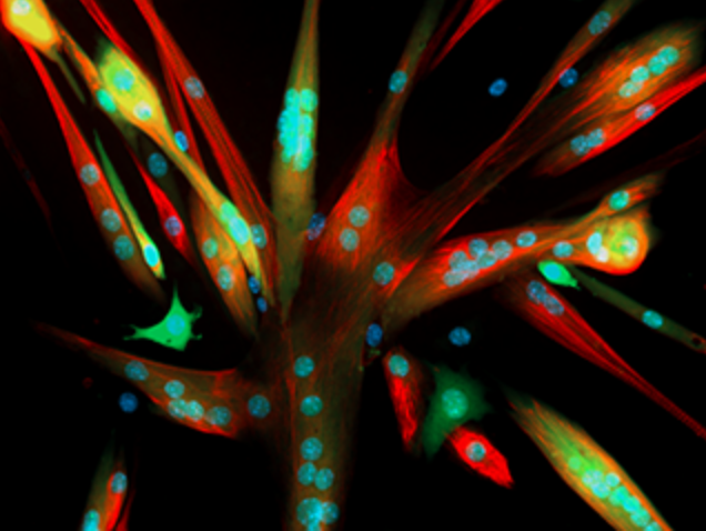
A fat signaling system controls the interface of cellular endoplasmic reticulum (ER) with early endosomes to regulate the shape and activity of mitochondria, and allow cells to survive periods of starvation, claim a group of scientists from FMP (Leibniz-Forschungsinstitut für Molekulare Pharmakologie), in Germany.
The findings were published in the journal Science on December 15, 2022 “Endosomal lipid signaling reshapes the endoplasmic reticulum to control mitochondrial function.” Fluctuations in access to nutrients is known to change the shape and function of intracellular organelles but how such alterations are coordinated across the cellular landscape has been unclear.

“We have found a completely new mechanism for how different compartments in the cell communicate with each other such that cell metabolism adapts in response to food supply,” said Volker Haucke, PhD, a scientist at FMP and the senior author of the study.
“This elegant study highlights the incredible complexity of intracellular processes and their continuous reciprocation with the environment. The researchers’ use of an ‘organelle conveyor belt’ analogy emphasizes that cellular activity is a form of internal engineering with seamless feedback among participating intracellular structures,” noted physician and evolutionary biologist, William Miller, Jr., MD, author of Bioverse (Miller was not part of this study).
While studying a rare genetic disorder, XLCNM (X-linked centronuclear myopathy), the investigators found that the gene myotubularin1 (MTM1)—a phosphatase enzyme that removes phosphate groups from specific lipids and that is mutated in XLCNM—regulates lipid turnover in early endosomes. This reshapes ER which in turn controls mitochondrial morphology and function.
XLCNM is a developmental disorder of skeletal muscles where muscle weakness affects movement and even breathing. Individuals with mutations in MTM1 do not survive beyond 10 to 12 years and in severe cases, die soon after birth.
“Muscles are highly sensitive to starvation. Their energy reserves are soon depleted. We therefore began to suspect that the defect in cells from XLCNM patients might be related to an incorrect response to starvation,” said Haucke.
When healthy cells are starved of nutrients, endosomes recruit MTM1. This prevents the phosphorylated lipid PI(3)P (phosphatidylinositol 3-phosphate) from establishing contacts (focal adhesions) between tubular ER membranes and early endosomes. The lack of ER-endosomal contacts converts ER tubules into sheets, blocks the division of mitochondria (mitochondrial fission) and maintains oxidative metabolism by burning stored fat.
In healthy cells, the ER forms a meshwork of flattened sacs near the nucleus of the cell and narrow tubules in the cell periphery. When healthy cells are starved the outer narrow tubules regress change into flat sacs. This altered structure of the ER enables the mitochondria to fuse together.
“Such greatly enlarged ‘giant mitochondria’ are much better able to metabolize fats,” said Wonyul Jang, PhD, lead author of the study.
The researchers observed, in cells deficient in MTM1, ER do not form flattened sheets and their membranes develop holes. Fats cannot be transported or burned efficiently in cells lacking MTM1. While in healthy cells, starvation reduces contact between endosomes and the ER, allowing the ER to change shape, in cells lacking MTM1, the contact between endosomes and ER is not reduced.
The endosomes pull on the ER, stabilizing the peripheral tubular ER structure and creating holes in its membranes. Peripheral ER tubules are responsible for mitochondrial fission. Therefore, in the absence of MTM1, the mitochondria remain small with limited capacity to burn fats, creating severe energy depletion in the cell.


