June 15, 2017 (Vol. 37, No. 12)
Researchers Find New Ways to Bring Microfluidic Devices into Labs and To Point of Care
The market for microfluidics has experienced tremendous growth over the past few years. Lab-on-a-chip devices for use at the point of care are allowing faster and more portable testing. Other microfluidic devices, for controlling and manipulating sub-microliter volumes of liquid in the laboratory, are saving researchers from needing complex, expensive equipment and highly trained staff.
Although microfluidics has huge promise, this remains an emerging field. There are challenges to the uptake and large-scale commercialization of these technologies—both in laboratories and at the point of care. Overcoming these barriers and developing novel approaches to this technology was discussed at the Selectbio Ninth Annual Conference on Biosensors, Microfluidics, and Lab-on-a-Chip Technologies, which was held in May in Munich.
One challenge to the commercialization of microfluidic devices is ensuring they’re consistently useful, affordable, and function properly. Jens Ducrée, Ph.D., professor of microsystems, Dublin City University, compares successfully scaling up microfluidic devices to selling mobile telephones. Customers expect their mobile to be relatively cheap and sufficiently well manufactured that it won’t have major flaws.
No Killer App
He explains that microfluidics doesn’t yet have a “killer app” that would automatically lead to high production numbers. Instead, companies need to streamline the way they develop new systems, somewhat akin to how in the 1980s and 1990s the development of integrated circuits brought computers into the home.
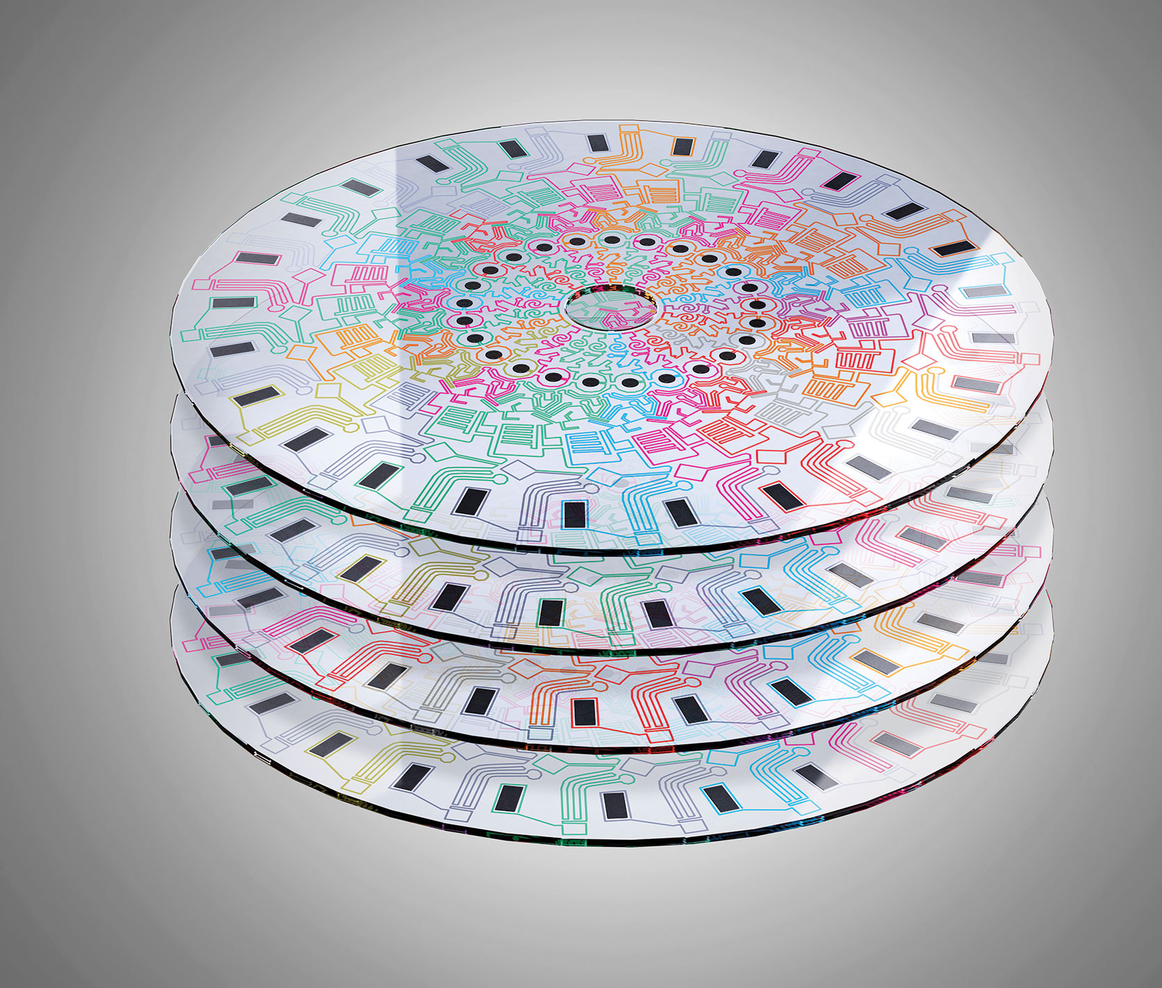
At the beginning of this year, Dr. Ducrée received several million euros of funding to commercialize a centrifugal lab-on-a-disc platform, a microfluidic system whereby liquids are propelled and manipulated by rotating a disc to induce a centrifugal force. “We have a streamlined approach,” he says. “We combine elements from a library of unit operations that represent typical operations in a lab, such as mixing or aliquoting.”
According to Dr. Ducrée, each unit in the library represents an operation, such as a resistor, capacitor, or diode in microelectronics. By combining these standardised elements, rather than developing new components for each application, it’s possible to quickly develop different microfluidic systems.
The main innovation of this lab-on-a-disc platform is event-triggered flow control where a valve opens when a liquid arrives at a defined location. The valve opening can trigger a string of operations, such as the release of wash reagents, elsewhere on the disc. According to Dr. Ducrée, this makes the system robust to manufacturing errors and disc handling, and makes it easier to set up a long chain of operations.
“In a platform approach similar to the automotive industry, we generate a variety of bioanalytical applications by combining elements from a parametrized library of unit operations that represent typical operations in a lab, such as mixing, metring, or aliquoting,” noted Dr. Ducrée.

At Dublin City University, scientists affiliated with the Biomedical Diagnostics Institute are developing centrifugal microfluidic lab-on-a-disc technologies. For example, these scientists are expanding the centrifugal microfluidic toolbox by introducing event- and pulse-triggered valving schemes that can establish logical, Boolean-type relationships. By combining different unit operations, the scientists demonstrate how to achieve higher-level integration and automation of various multistep, multireagent, multiparameter test panels for nucleic acid amplification, immunoassays, and cell counting.
Accelerating Microfluidic Chip Fabrication
Another challenge to commercialization is speeding up the fabrication of microfluidic chips. Chips are often manufactured at a wafer level, but the final processing steps involve laborious chip-by-chip processing. For example, a high-speed rotating saw is used to dice the wafer and this is cooled with a water jet. The micro-particles thrown off by the saw can contaminate chips that are pre-integrated with biological reagents, and the water can disperse the reagents.
Emmanuel Delamarche, Ph.D., research scientist, Zurich Research Laboratory, IBM, helped develop a new microfabrication process where the chips are wholly fabricated at a wafer level. After the electrodes and microfluidic channels are cut, the wafers are partially diced, cleaned, and dried. An ink-jet spotter places the biological reagents and then the wafer is laminated at temperatures as low as 45°C.
“The wafers are pre-diced so the user can simply break off a single chip,” he explains. “We call it ‘chip-olate’ because it’s like breaking a chocolate piece from the bar.”
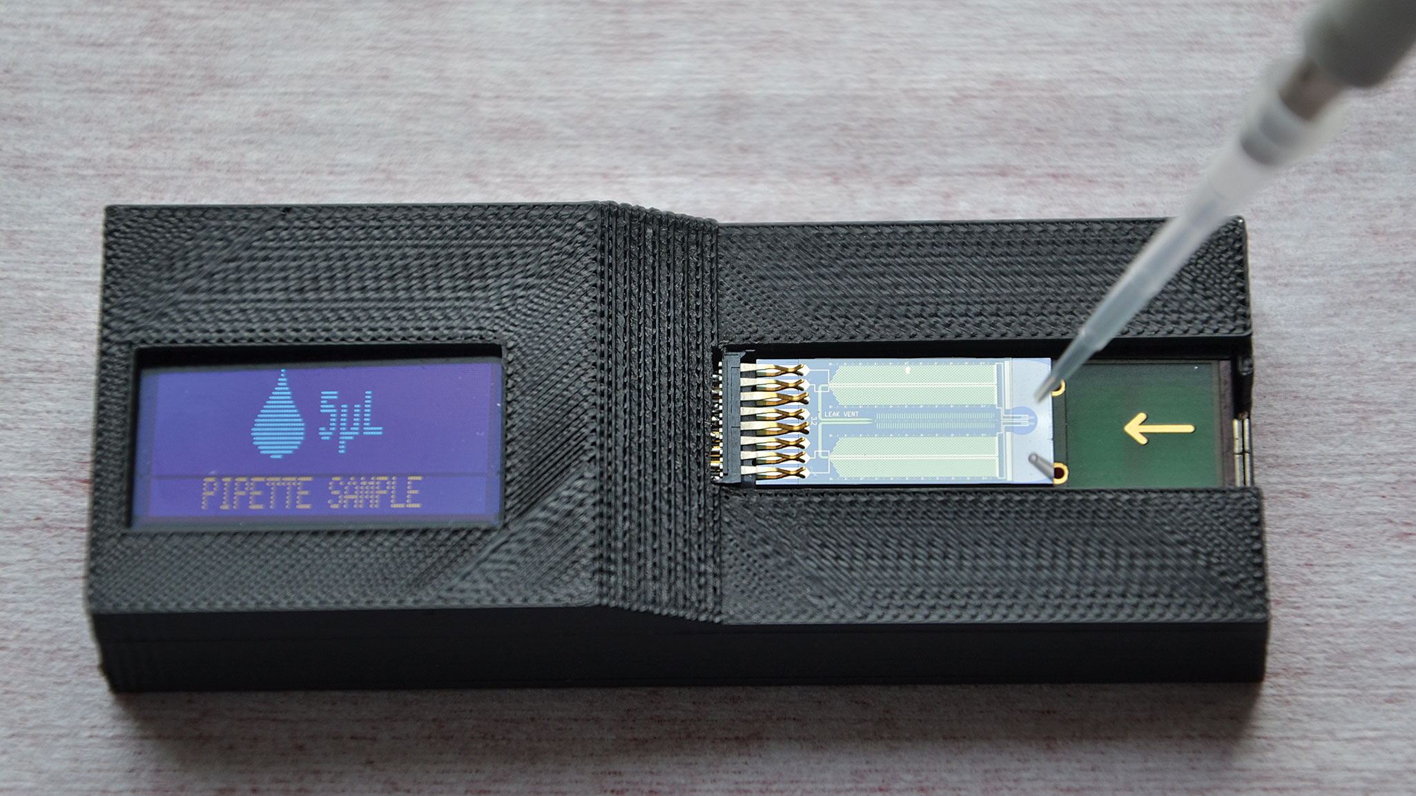
Dr. Delamarche also talked about adding microelectrodes to microfluidic chips so the fluid flow can be monitored by a smartphone in real time. He explains that information about fluid leaks, viscosity, or anomalies can be followed with sub-nanoliter precision to aid calibration of diagnostic tests and to flag up errors during testing. “In point-of-care diagnostics, if something goes wrong with a test, it wastes precious time for the patient,” he notes.

IBM’s Zurich Research Laboratory is working to extend the performance of microfluidics for point-of-care diagnostics. The microfluidic chip in this image not only integrates modular capillary-driven elements, it also contains microelectrodes that monitor capillary flows with submicroliter precision. When the chip is plugged into a Bluetooth-enabled peripheral, as it is here, it can communicate flow information to a smartphone in real time.
Light and electron microscopy have made huge advances in the past few years. In 2014, for example, the Nobel Prize was awarded for using fluorescent molecules to circumvent a long-time limitation of optical microscopy—imaging objects smaller than the wavelength of light.
Since light and electron microscopy are highly complementary techniques, it would often be ideal to combine both to study cell structures and dynamic processes, such as membrane trafficking or the dynamics of organelles. The problem with achieving this is that samples need to be rapidly frozen to a very low temperature to get the best resolution with electron microscopy. Cellular processes, meanwhile, can happen within seconds or milliseconds.
According to Thomas Burg, Ph.D., group leader, Max Planck Institute for Biophysical Chemistry, this has made it hard to study some biological processes with light microscopy and use electron microscopy to look at cell structures at the same time. “Water doesn’t like to cool fast,” he says. “And the methods of cooling have remained almost unchanged since the 1960s.”
Dr. Burg has helped develop a microfluidic device to rapidly freeze samples for cryomicroscopy during live imaging. The sample is placed inside of a thin polymer foil, 50 micrometers thick, above a thin electrical heater. Below that is a thermal insulator and then a liquid nitrogen heat sink.
“It’s a completely new concept in microfluidics,” he says. “You can get dynamic imaging almost instantaneously with cryofixation. The microfluidics allows the user to choose a time point for imaging with millisecond precision because the small size allows rates of cooling necessary to prevent the formation of crystals in ice.”
The heater can be turned off at a desired time, and the microfluidics cools on command extremely rapidly because the nitrogen is very cold and the small device operates far from thermal equilibrium.

A technology developed by the Max Planck Institute for Biophysical Chemistry enables high-quality structural preservation of biological samples by microfluidic cryofixation. Samples may be prepared in a light microscope and with millisecond time resolution. Because the technology achieves ultrarapid freezing, it prevents the formation of ice crystals that can disrupt cellular structures. This technology may bridge live-cell imaging and postfixation ultrastructural techniques such as electron microscopy.
Simplifying Processes
There are several devices on the market for emulsifying samples for DNA amplification techniques, such as digital polymerase chain reaction (dPCR), which is PCR performed on tens of thousands of nanoliter-sized water-in-oil droplets, which each act like a tiny test tube or well. The trouble is that, with current technology, multiple handling steps and specialist machines are needed.
Felix von Stetten, Ph.D., associate director, Hahn-Schickard, has helped develop a microfluidic unit operation that can emulsify, amplify, and detect DNA in a single cavity. The technology uses centrifugal forces to carry the sample from an inlet chamber to a nozzle where droplets are formed. Within the same microfluidic chip, the droplets are incubated for DNA amplification and readout performed with a microarray reader.
“The novel thing about this technology is that everything is integrated into a single chip,” he says. The chip can be
either processed as a single processing device or with standard laboratory devices. The novel technology can be used to perform digital PCR (dPCR), digital recombinase polymerase amplification (dRPA), and loop-mediated isothermal amplification (LAMP).
One innovative application of the technology is encapsulating bacteria, such as MRSA, into droplets and performing dRPA on individual cells. “This allows you to analyze whether a species gene and an antibiotic resistance gene are contained within one cell, and to select the appropriate antibiotic,” Dr. von Stetten explains. Performing dRPA on the bacteria before distributing DNA to the droplets can shear the genome, so it would be unclear which bacterial species have which resistance.
“I’m somewhat biased, but I’d say the areas where microfluidics will play a huge role in diagnostics is very simple devices,” says David Weitz, Ph.D., Mallinckrodt Professor of Physics and Applied Physics, Harvard University. He adds that devices that use valves are often expensive, difficult, and less commercially successfully. “I think the next developments are in drops.”
At the conference, Dr. Weitz presented on a high-throughput approach for barcoding
individual cells by capturing them in nanoliter droplets. The cells are captured along with hydrogel microspheres carrying barcoded primers and reverse transcription reagents. After encapsulation, the primers are released and tag RNA during reverse transcription reactions. The droplets are then broken, the nucleic acids collected, and processed using established techniques as a normal sequencing run.
“It’s remarkably simple in the way it works,” says Dr. Weitz. “And it’s the simplicity that gives it power.”
Dr. Weitz has used droplets to barcode mRNA from thousands of mouse embryonic stem cells. The technique may, in the future, be used to map other molecules involved in gene expression, such as proteins. This can help researchers understand physiological events, such as the cell cycle, aging, and infection at the level of the individual cell.

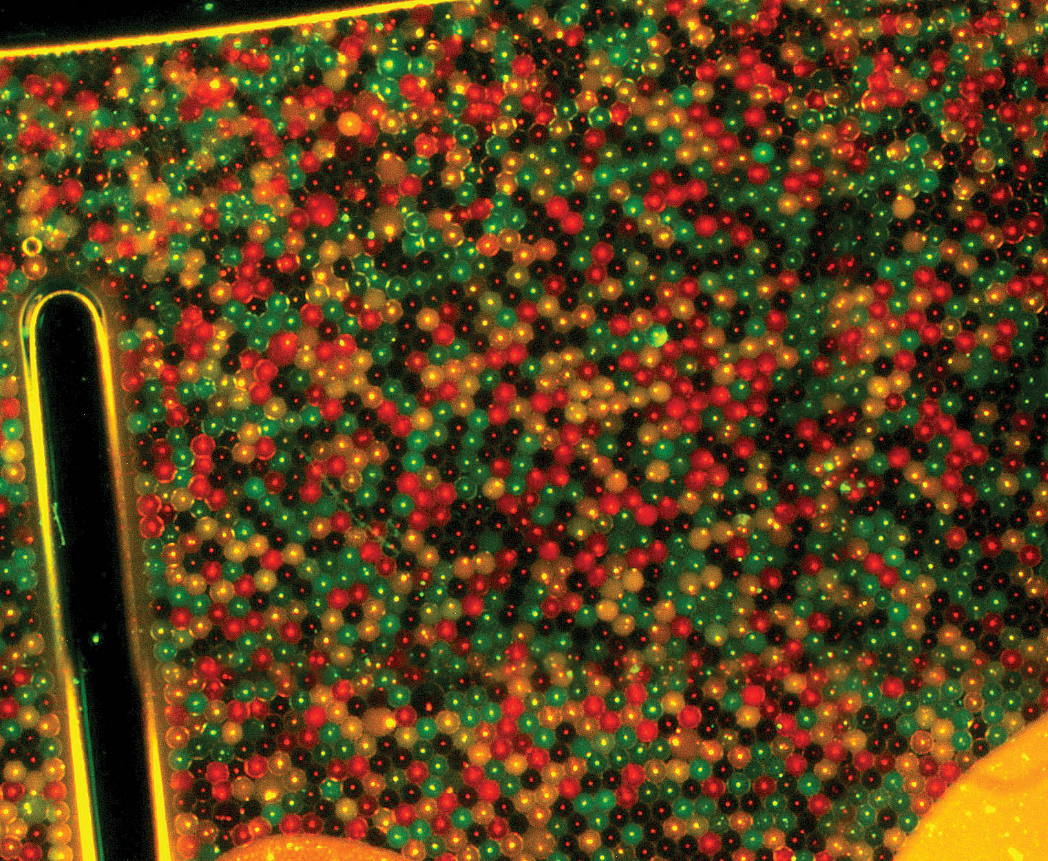
Digital recombinase polymerase amplification (dRPA) was used in multiplex (biplex) mode by Hahn-Schickard scientists who are participants in a project called IDAK—isothermal digital single-cell amplification for the detection of antibiotic-resistant pathogens in hospitals. IDAK is building on dRPA technology from Hohenstein Institut für Textilinnovation to develop a fast and mobile diagnostic system. This image depicts how multiplex dRPA can detect pathogens and determine their resistance capabilities at the same time. The analysis of droplet-encapsulated bacteria reveals vicK, a gene unique to Staphylococcus aureus (red); mecA, a resistance gene (green); both genes (orange); and negative results (black).


