May 15, 2014 (Vol. 34, No. 10)
XF Bioenergetic Analysis Identifies Defects in Human Skin Fibroblasts
Mitochondrial disorders are characterized by defective mitochondrial function in metabolically active tissues such as skeletal muscle and brain. While spectrophotometric analysis can be utilized to detect biochemical defects in muscle biopsies or skin fibroblasts, samples are difficult to obtain, require a large number of cells, and this method cannot detect defects in the interplay between complexes and subunits of the oxidative phosphorylation system.
In addition, mitochondrial disorders are heteroplastic, with only a fraction of mitochondria affected, and so require a more sensitive method for analysis.
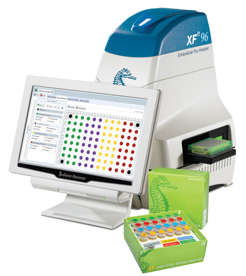
The Seahorse Bioscience XF96 Extracellular Flux Analyzer (Figure 1) offers the advantage of significantly increased sensitivity, enabling the detection of defects in the OXPHOS system in human skin fibroblasts. In contrast, traditional methods results with skin fibroblasts often do not coincide with results from muscle biopsy-derived material. The example below describes an assessment of the XF96 Analyzer’s ability to measure mitochondrial defects in human skin fibroblasts.

Figure 1. XFe96 Extracellular Flux Analyzer and XF Cell Mito Stress Test Kit
XF Bioenergetic Analysis
Oxygen consumption rate (OCR) and extracellular acidification rate were measured in adherent fibroblasts with the XF96 Analyzer, 24 hours after seeding in the XF96 cell culture microplate (Figure 1). The drug injections ports of the XF96 Assay Cartridge were loaded with the assay reagents at 10X in assay medium. 20 μL of oligomycin (10 μM), and 22 μL of FCCP (7 μM) were added to ports A and B respectively.
Culture medium was exchanged with assay medium prior to measurements. Culture medium was aspirated and 180 μL prewarmed PBS added, aspirated and 180 μL prewarmed assay medium added. 180 μL of assay medium was added to the temperature control wells (without cells), and the microplate equilibrated in a CO2-free incubator at 37ºC for 30 minutes.
During this equilibration period, the XF96 Analyzer was calibrated using a calibration plate and the standard XF calibration protocol. Following calibration, the plate was replaced with the XF96 cell culture microplate containing fibroblasts and the skin fibroblast assay started. The resulting data was analyzed using XF software.
Interpretation of Results
The assay described is a version of the XF Cell Mito Stress Test, which reveals key parameters of mitochondrial function: basal respiration, ATP production, and respiratory capacity. The XF Cell Mito Stress Test measures the basal oxygen consumption rate (OCR, reported as pmol per minute per cell) to obtain the basal respiration—or resting state of the cells. A decreased basal rate in patient vs. healthy control samples may indicate a defect in the respiratory complexes.
In this study 17 patient samples were tested. Using traditional spectrophotometric methods, nine of the seventeen patient fibroblast samples were found to have a genetic defect, while nearly half falsely tested normal. Analysis of mitochondrial-enriched homogenate from muscle yielded two false negatives (normal).
Upon analysis of the skin fibroblasts using the XF96 Analyzer, all 17 yielded results indicative of a defective OXPHOS system, demonstrating the value of analyzing the intact OXPHOS system rather than individual complexes, and the value of the increased sensitivity of the XF96 Analyzer. This assay also utilized the XF Cell Mito Stress Test, which measures the basal OCR to obtain the basal respiration.
Research suggests that decreased basal rate in patient vs. healthy control samples may indicate a defect in the respiratory complexes (Figure 2). Oligomycin blocks ATPase activity, and the reduction in OCR is a measure of the respiration needed to sustain ATP consumption in the cells. The remaining respiration reflects the proton leak of the mitochondria. FCCP uncouples respiration by carrying protons across the IMM, and FCCP also dissipates the electrochemical gradient (membrane potential) that drives ATP synthesis.

Figure 2. Diminished spare respiratory capacity (SRC) in patient skin fibroblast samples, relative to the corresponding control samples
The results (Figure 3) demonstrated that an optimized concentration of FCCP reveals the maximal respiration (reaction to increased ATP demand). Reduced maximal respiration may lead to an energetic crisis for the cells. Because mitochondrial diseases are often heteroplasmic, the “healthy” fraction of mitochondria will be able to increase their respiration to the same degree as the healthy control samples upon FCCP stimulation. Thus, looking at fold changes may give the impression of both populations being healthy, whereas using the XF Analyzer and viewing data normalized to the number of cells per well reveals the deficient maximal respiration.

Figure 3. Mitochondrial stress profiles of healthy (pink) and patient (brown) fibroblasts. Oxygen consumption rate (OCR) measured in human skin fibroblasts derived from either healthy control, or patient with defined DNA mutation, in an OXPHOS gene.
Summary
The advantages of using the XF bioenergetic assay for measuring bioenergetic dysfunction in human skin fibroblasts are twofold. First, the intact oxidative phosphorylation machinery is being measured, allowing detection of mutations affecting the interplay between complexes and subunits of the respiratory chain. Second, only a very small amount of clinical material is required and sensitivity is greatly enhanced. Thus, defects that could not previously have been detected in a reliable way using skin fibroblasts are now detectable through bioenergetic analysis, suggesting that skin fibroblasts may be used as a first line of screening for suspected mitochondrial disease.
The greatly enhanced reliability of detection allows the results to be used as justification for the more invasive sampling of muscle biopsies for further analysis. In the future, it is expected that methods will be developed describing the analysis of permeabilized cells, enabling researchers to determine which complex is affected.
Per Bo Jensen, Ph.D. ([email protected]), has worked as a research scientist at Seahorse Bioscience. Dr. Jensen is currently working as a research scientist at Novo Nordisk in Copenhagen.


