October 15, 2016 (Vol. 36, No. 18)
One of the major roadblocks to curing cancer is modeling the complexities of the disease; thus, increasingly refined models of the disease are necessary.
The result has been a sizable increase in the number of genetically engineered and, more recently, humanized animal models for use in research and preclinical studies.
Using advanced genetic techniques such as clustered regularly interspaced short palindromic repeats (CRISPR) and transcription activator-like effector nucleases (TALENs), scientists have created various immunological knockouts that reduce the obstacles intrinsic to xenografting human immune systems and human tumors. These models, well established in mice, are also being developed in rats, swine, and other animal models.
At EMD Serono, a biopharmaceutical division of Merck, patient-derived xenograft (PDX) models of cancer are being used in all stages to support clinical decision making. “For all of the PDX models,” says Anderson Clark, Ph.D., director of translation in vivo pharmacology, at EMD Serono, the North America biopharmaceutical business of Merck KGaA “immunocompromised mice are utilized.” EMD also uses cell-line xenografts, called CLDX, in other applications, but believe that data from PDX models translates better into the clinic.
“Tumors from PDX models display complex genomic expression profiles and histological characteristics that are similar to the patient tumors from which they are derived,” says Dr. Clark. “We use these models to choose indications and combination partners for our oncology drugs and to identify response markers and responsive patient subpopulations.”
However the cost associated with patient-specific modeling is still high. Merck made a decision in 2010 to build this platform using models from academic collaborators and CROs because of its probability of success in helping patients, and has already expanded from supporting Phase II clinical trials to also supporting Phase III trials. “The next big growth area for PDX models,” he says, “will likely be for testing immuno-oncology treatments in humanized mice.”
According to Michael Seiler, Ph.D., portfolio director for commercial genetically engineered models at Taconic Biosciences, “The publication history for humanized mice dates all the way back to the early 1980s. But as we know, the acceptance and general utility of humanized animal models for preclinical efficacy in drug discovery can take a significant amount of general acceptance in the scientific literature before it crosses over. I think we are starting to see that emerge.”
Early work on mouse humanization was accelerated by the generation of the NOG mouse, developed by the Central Institute for Experimental Animals (CIEA) in Japan (from which Taconic gets its exclusive license for its immunodeficient animal).
“The NOG mouse was a combination of the unique background of the nonobese diabetic (NOD) strain of mouse combined with the spontaneous scid (severe combined immune deficiency) mutation, which resulted in attenuation of the immune system,” says Dr. Seiler, “The real magic happened when the CIEA crossed the scid mouse with a targeted mutation of the gamma chain of IL-2 receptor (IL2rg).” The combination of one spontaneous mutation (scid), one targeted mutation (IL2rg), and the NOD background strain results in the severe immune deficiency of the NOG mouse.

Taconic exploits this in the development of its humanized mouse models program. Their latest version, called huNOG-EXL, takes advantage of the NOG mouse immune deficiency and builds upon it with transgenic expression of critical human growth factors to stimulate more complete immune system development. Cells from the human innate and adaptive immune system are developed, essentially replacing some of the missing components of the host mouse immune system. The immune-deficient host creates a niche for the human cells to take up residence, and growth factor expression solves one of the major limitations in human immune cell development.
This combination is an extremely powerful tool for immune-oncology pharmaceutical testing. “There are two main things you can capture in the humanized mouse,” says Dr. Seiler. “First is the efficacy of treatments activating an immune response against the tumor burden itself in a combined human xenograft/immune systems xenograft strategy. But the other, more important, aspect is that you can capture the mechanism of action of a uniquely specific humanized monoclonal antibody drug on a human cell type.”
Developing the Rat Model
Mice have been used because of increased availability of immunodeficient models; however, one company, Hera BioLabs, has begun work to humanize the rat. “To our knowledge, we are the furthest along in developing humanized rats for commercial use,” says Tseten Yeshi, vp of research and development, which based its humanization efforts on the double Rag2, interleukin-2 (IL2) knockout (scid) rat. Like the mouse, the removal of the endogenous immune system is the first step in the humanization process.
“We started work on humanizing the liver,” said Yeshi. “It’s a two-step process once one has the suitable immunodeficient model. First we ablate the endogenous liver of the animal to a certain percentage, and then we repopulate that ablated liver with human hepatocytes.” Depending on the purpose, between 25% and 75% of the liver must be ablated before being reseeded with human cells, typically human primary cells. Hera also plans on using other commercially available hepatocyte-like cells.
The animal model field is highly competitive, says Yeshi. “Well-established players like Jackson Laboratories and Taconic have a huge focus on humanized models.” Rats, he points out, are the primary rodent model used in toxicology studies, “because it has a lot of advantages over the mouse, such as favorable absorption, distribution, metabolism, and excretion/pharmacokinetics (ADME/PK) characteristics, ease of handling, ability to sustain more intense intravenous dosing, increased frequency of blood sampling with increased sample size, and larger tissue samples.”
Hera has been in existence for just over one year and is currently providing human cancer xenograft and PDX efficacy and creation services in the scid rats as it develops the humanized models. “We are in the optimization and validation stages right now,” says Yeshi. “We have to look at how much humanization we can achieve in the rat liver. Following that, we need to run some preliminary studies showing human-specific drug metabolism in these rats so that we can demonstrate the effectiveness of the model.” The next goal will be the humanization of the immune system in the rat as well.
“Immuno-oncology is a huge field, and so interactions of particular drugs and compounds with the human immune system have a big impact on discovering new drugs.” Hera aims to be the first to accomplish this in rats.

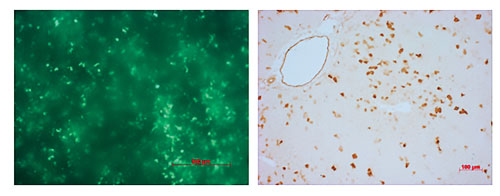
The humanization of the liver and immune system in rat models is underway at Hera BioLabs, which provided this image depicting rat liver gene delivery for the purpose of endogenous liver ablation, as measured by GFP expression. GFP was delivered to the liver of neonatal rats, and the liver was analyzed at two weeks. Left: Fluorescence in a whole liver lobe. Right: Immunohistochemistry of a liver section using an antibody that recognizes GFP.
Advantages of the Swine Model
While smaller animals provide for quicker study turnaround, larger models are closer in complexity to the human population. David Largaespada, Ph.D., is a professor of pediatrics and associate director of basic science at the Masonic Cancer Center at the University of Minnesota and CSO for Surrogen, a gene-editing company working to develop humanized swine models and models of complex diseases.
“What we have done,” says Dr. Largaespada, “is generate a swine model of neurofibromatosis type 1 syndrome, or NF1.” NF1, also known as Recklinghausen’s disease, is a genetic disorder involving the growth of multiple benign tumors called neurofibromas. The goal here is to create an animal that replicates some of the features of the disease that don’t occur in mice, such as external neurofibromas, which are hallmarks of NF1 syndrome. Furthermore, devices used in the treatment of diseases in humans cannot be tested well in small animal models.
Pigs, such as the Ossabaw miniature pigs used at Surrogen, grow to around 180 to 200 pounds, similar in size to humans. Finally, it has been shown that NF1 patients metabolize drugs differently than control populations. “We would like to use the pig models to do pharmacology studies,” says Dr. Largaespada, “where we’re testing some of these drugs in NF1 swine and their (control) litter mates.”
Broadly, Dr. Largaespada and his team hope to develop swine models for a variety of human diseases with the largest public health impact, including metabolic syndrome, Alzheimer’s disease, and cancer. They have succeeded in creating an immunodeficient pig to rival the rodent models as well. “Of course these diseases are complex,” he says, “and engineering the right, relevant, genetic changes into the pig isn’t a simple thing.”
Surrogen, a wholly owned subsidiary of Recombinetics, uses an array of molecular approaches to mutate large animal genes; the company has a license to use TALENS, for example, in pigs and cows. One approach is to create humanized versions of certain pig genes, says Dr. Largaespada, “i.e., replacing them with the human version.” Surrogen is in dialogue with academic and industry partners interested in reliable models for testing drug efficacy against human proteins.
Modeling Complex Workflows
As animal models become more complex, the systems used to track preclinical animal study data need to keep pace. One company focused on simplifying and standardizing workflow is Studylog Systems, which began 13 and a half years ago in Eric Ibsen’s garage. “I had been running a research group at a Bay Area biotech company and was really struggling to manage the workload and data generated, using Excel macros, Microsoft Outlook tools for scheduling, PowerPoint presentations, and Prism files,” says Ibsen, vp and co-founder.
“Those disparate file types were all part of a study folder usually accessible to just the originator,” he recalls. “A year or two later you had to go into the folder and look at the studies. It was really challenging trying to recall anything.” A workflow tool built on top of a database, “so that you could search and find things and compare across studies,” would solve this problem.
Ibsen and his collaborator created Studylog Systems, a program for investigators that allowed for the running of preclinical animal studies in a way that mirrors clinical trials, increased throughput, and preserved the detailed data for future use. This automation of workflow, including the study design, planning, and data analysis, greatly reduced the personnel and resource costs associated with tracking animal studies.
Once the study is set up by the various stakeholders, users are notified and data collection can begin. The animals are put into groups through manual or randomized processes, and Studylog automates the collection of measurements, samples, and the performance of tasks. Once data is recorded, users can highlight individual studies and create Excel outputs, graph the data, or analyze it with internal and external statistical packages.
“We have automated and standardized an inherently variable process,” says Ibsen. “It’s something that Excel-based Lotus-Intel-Microsoft (LIM) systems have heretofore been unsuccessful in being effective in because they don’t reflect the workflow, nuances, metadata, and differences from study to study.”
Beginning with oncology-based systems, Studylog has since expanded its software capabilities into other animal models. “We are used by seven out of ten big pharmas in oncology,” Ibsen says, “and there are several, including the largest biotech companies, that have deployed us worldwide in all disease areas.”
The value of his animal study management system is going to be even more important as the industry moves into individualized models of disease, such as PDX, he says. “The rate limiter is that doing staggered staging enrollments,” which will become more prevalent in individualized disease models, “is just too labor intensive at any given time for most labs. We are changing the game because we make it easy to randomize animals iteratively and easily collect and align their data.”
PDX Models
Despite great progress, most newly developed anticancer agents fail clinical trials, mainly due to the use of inappropriate preclinical models. In this context, patient-derived-xenograft (PDX) models regained interest over the last decade because, compared to cell lines, they better preserve cellular heterogeneity of the original patient tumors and allow investigation in solid tumor with a microenvironment. They also more faithfully mimic clinical response than commercial cell lines.
To improve the translation of preclinical to clinical results, Charles River Laboratories (CRL) developed a collection of more than 450 PDX models for compound evaluation, which covers the diversity of cancers.
“This collection can be used to evaluate drug performance by using ex vivo 3D clonogenic assay platforms,” says Vincent Vuaroqueaux, Ph.D., who heads the biomarker development and bioinformatics department at CRL in Freiburg, Germany. “This approach offers a unique opportunity to evaluate compound efficacy and selectivity in a heterogeneous cellular context and to identify the most sensitive tumor subtypes.
“By comparing ex vivo drug sensitivity results to the whole molecular characteristics of models, PDX allow identification/validation of predictive biomarkers of clinical utility at an early stage, which is essential for better cancer patient enrollment into clinical trial.”
CRL’s PDX also helps to improve the translation of preclinical to clinical results by allowing compound evaluation in vivo, continues Dr. Vuaroqueaux, adding that PDX in vivo testing is a decisive step prior to clinical trial, to work on dose findings, to address the microenvironment, drug delivery and side effects.
The development of a mouse clinical trial helps in forecasting responses observed in clinical trials by assessing compound sensitivity across tumor genetic diversity to define appropriate schema for subsequent clinical trials and to validate biomarkers for models/tumor selection.
“Altogether, CRL PDX helps in preparing clinical trials and in selecting the right tumors and the right biomarkers,” notes Dr. Vuaroqueaux.
Ian Clift Ph.D. is a Scientific Communications Consultant, Biomedical Associates and Clinical Assistant Professor, Indiana University


