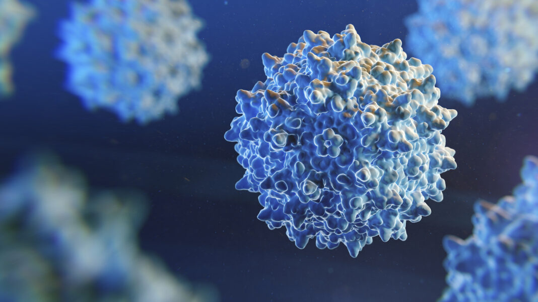
Separating full and empty adeno-associated virus (AAV) capsids during the manufacture of single-gene therapies remains a significant challenge. Ultracentrifugation and anion exchange chromatography (AEX) are common differentiation methods, but their success is, respectively, unstainable at scale and serotype dependent.
Distinguishing between full and empty capsids based upon AAV structural changes when the capsids envelop a gene could be an alternative, according to Caryn Heldt and Molly Skinner, both at Michigan Technology University, and Ganesh Anand at Pennsylvania State University, writing in Biomedicines.
“It has long been thought that the charge difference between empty and full AAV was because the highly negative core (e.g., the DNA inside of the capsid) would impart a negative charge to the surface of the capsid,” Heldt tells GEN. “However, proteins have no dipole moment, so charge should not transmit through the capsid.”
They propose four reasons the structure of AAV could change depending on whether or not the capsid contains DNA.
“One possible change could be that capsid expansion may correlate with being filled,” Heldt says.
“The increase in packaged genome size increases the internal pressure of some viral capsids,” she and her team point out, which enlarges the capsid and, therefore, changes its conformation. “This is the only theory that has been shown in multiple AAV serotypes as creating a possible difference between empty and full capsids. This is especially relevant for AAVs… that use the metabolic energy of the host to insert DNA into a preassembled capsid,” rather than condensing around the genome.
In making that determination, Heldt and colleagues also investigated changes in surface chemistry as a possible differentiator. When a capsid interacts with negatively-charged DNA, it may contract and bury some of the surface amino acids. To this point, they show that an empty AAV5 capsid is approximately 6 nm larger in diameter than a full capsid containing the GFP gene. Changes in an AAV8 capsid, however, were quite small.
They also explored the possibility of linking capsids’ status to whether the N-tail of virus particles is tucked into the five-fold pore in the presence of DNA. However, “tail-tucking may not be the case for AAVs,” Heldt’s team reports, and the N-tail may be inside the capsid. For this approach to be useful, the N-tail structure must be known.
Finally, the scientists consider the quantity of the structural protein VP3 on the capsid’s surface, observing that the surfaces of full recombinant AAVs contain more VP3 than the surfaces of empty ones. However, because the ratio of capsid proteins in the final AAV structures is somewhat random, this is not yet a reliable way to determine whether capsids are full or empty.
Using these insights, potential separation techniques may include using high pressure to deform empty capsids, or using high-pressure filtration to squeeze malleable, empty capsids through membranes.
“I hope this paper will give the community more ideas to explore how conformational changes on the capsid may be the cause of the charge differences between empty and full AAV capsids,” Heldt says.


