June 15, 2013 (Vol. 33, No. 12)
Simple Cell-Based Monitoring System Aims to Pack Twice the Punch Inside Incubators
Cultivating cells may take days to weeks, making it difficult to monitor morphological changes. The long-term time-lapse monitoring of cells can provide reliable data about cell growth and death, and morphological changes. Cell proliferation and death measurement is a basic function in drug toxicity and safety testing, as well as in biotherapeutic research.
Continuous monitoring of cells in the same environments may require environmental regulators as well as monitoring systems. A high-quality microscope and incubator simply cost too much to use just to check for morphological changes of cells and their confluence. Unfortunately, until recently there were no compact microscopes that could be easily placed inside a thermo-hygrostat.

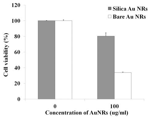
Figure 1. Cell viability test of SH-SY5Y: Silica-coated and Bare AuNRs were treated for 24 hours and viability of cells was measured using Cell titer-glo assay. Silica-coated AuNRs reduced toxicity compared to bare AuNRs.
NanoEnTek’s Juli™ Br can record live-cell images and fits in most incubators because of its compact size. One main board outside of the chamber can control two microscopes, monitoring two different experiments simultaneously. Using cost-effective dual Juli Br, we performed a simple experiment using SH-SY5Y (neuroblastoma) cells.

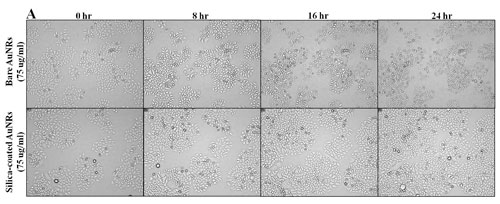
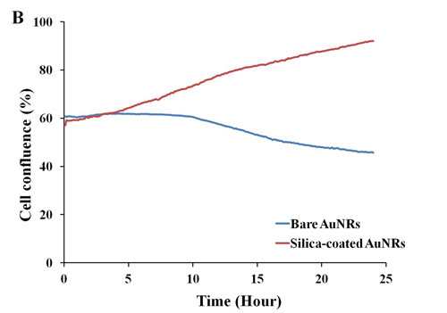
Figure 2A. Recording of SH-SY5Y cells: The cells gradually grew from confluence 57.17% to 92.10% when silica-coated AuNRs (75 µg/mL) were incubated. On the other hand, bare AuNRs (75 µg/mL) caused the cell death, which dropped from 60.75% to 45.73% confluence.
Toxicity Testing
Various applications of gold nanorods (AuNRs) were featured because of their photo-thermal properties in cancer therapy and drug delivery research. The surface of AuNRs can be modified to reduce toxicity. We used silica-coated AuNRs to increase biocompatibility. Nanoparticles are as small as subcellular components, which can result in membrane disturbance through its surface. AuNRs mostly existed in cytosols through endocytosis. Therefore, cell viability of SH-SY5Y exposed to gold nanorods for 24 hours was determined by Cell titer-glo assay.
The cells were incubated for 24 hours with bare or silica-coated AuNRs (100 µg/mL). In the presence of bare AuNRs, viability of SH-SY5Y was reduced to 33.85 ± 0.69%, while 80.24 ± 4.25% cells were live when silica-coated AuNRs were treated (Figure 1).

Figure 2B. Recording of SH-SY5Y cells: This curved graph shows fluctuation of cell confluences compared between bare and silica-coated AuNRs.
Juli Br recorded images of SH-SY5Y cells at five minute intervals, and confluence was also measured. The cells cultured in six-well plates were treated with 75 ug/mL of bare or silica-coated AuNRs (Figure 2A). Confluence data is shown in Figure 2B.

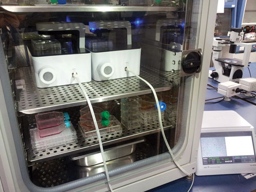
Figure 3. Juli Br (dual) installation in an incubator: Two microscopes can be controlled by the main device to calibrate focus and confluence measurement. They are compact enough to fit in most incubators without any problem. Incubator temperature does not affect the machine.
With simple experiments, we confirmed that bare AuNRs showed higher toxicity causing more cell shrinkage and membrane distortion than silica-coated AuNRs.
In this experiment, reduced nanotoxicity of surface-modified AuNRs was observed using Juli Br (dual). The two-microscope assembly took up to only one-third of the space in an incubator, making it easy to install in most cell culture laboratories (Figure 3). Small and easy-to-use Juli Br provides an easier way to monitor cells and measure confluence. A desktop program supplied with the machine can be utilized to edit videos.
Conclusion
Dual microscopes which can be controlled by one main device, are easy to use and easy to install inside an incubator. Juli Br (dual) can monitor the growth and death of cells from days to weeks. Videos clips generated from the time-lapse recorder can easily monitor cellular changes or migration.
Mino Kang, Jae Yeon Joo, and Seong Soo A. An are in the dept. of bionanotechnology, Gachon University, Gyeonggi, Korea.


