Doug Auld, Ph.D. Novartis Institutes for BioMedical Research
A literature review of a paper where the authors describe a method that can make FISH procedures more sensitive and specific.
This article* describes a strategy to improve the sensitivity and specificity of FISH procedures. The method employs a so-called padlock oligonucleotide that has ends that are complementary to the 5′ and 3′ of target sequences. Upon binding to single-stranded DNA (ssDNA) the probe circularizes (forming a loop between the two complementary ends), and if there are no mismatches in the hybridized region then a ligation reaction can be performed to close the loop. The ligation reaction is extremely sensitive to mismatches so even a single nucleotide mismatch will prevent ligation of the padlock probe ends.
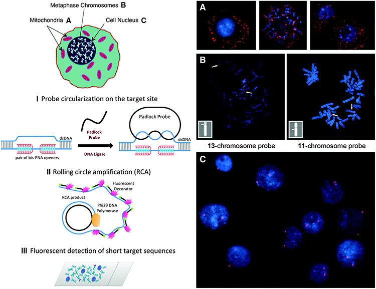
To make the target sequence ssDNA, the authors* used bis-PNAs (peptide-nucleic acid) openers that form both Watson-Crick and Hoogsteen base pairs with one strand of the target sequence thereby opening the double-stranded (ds)DNA. Finally once the padlock probe is ligated, a rolling circle amplification (RCA) reaction is performed which yields thousands of copies of the target sequence, and fluorescent-labeled oligonucleotides are then hybridized to the RCA product to generate the FISH signal (see Figure). The RCA reaction leaves the copies attached to the target sequence so these signals are visualized as punctate staining in fluorescent microscope images (see Figure).
Figure. Schematic depiction of the new method for sensitive and specific targeting of unique sequences on nondenatured human genomic DNA and representative images observed by fluorescent microscopy in experiments performed according to the scheme schematic. Our method consists of three major steps. (1) The PNA openers specifically bind to two closely located homopurine DNA sites that are separated by several mixed purine-pyrimidine bases and locally open the dsDNA. This opened region serves as a target for hybridization of an ODN probe to form a PD-loop. This binding is extremely sequence specific because nothing but the target location is accessible to the probe. (2) The assembled circular DNA serves as a template for the RCA reaction, which yields a long, single-stranded amplicon that contains thousands of copies of the target sequence. (3) For the detection step, fluorophore-tagged decorator probes are hybridized to the RCA product. The DNA amplicon remains firmly attached to its site of synthesis, so the multiply fluorescent-labeled product can be imaged as a visible point source that can easily be detected with a fluorescence microscope. (A) Multiple fluorescent spots in the vicinity of metaphase chromosomes and nuclei indicate the detection of short, human-specific DNA sequences within the mitochondrial genome in the cytoplasm. Data for target site MT-ND3 (5'-AAGAAGAATTTTATGGAGAAAGG-3') designed for the mitochondrial-encoded NADH dehydrogenase 3 gene are shown. (B) Two fluorescent spots are clearly seen on human chromosomes 13 and 11, identified cytogenetically, when probe (5'-GAGGGAGGTAGCCAGAGGAAG-3') specific to chromosome 13 or probe ALDH3B1 (5'-GAGGGAAGACCCAGGAGGGAGG-3') designed for the aldehyde dehydrogenase 3 gene on chromosome 11 is applied. Insert shows the location of these spots correlates to the position of the target sequence determined by Human Genome BLAT (http://genome.ucsc.edu/cgi-bin/hgBlat). (C) Typical results observed in the large majority of nuclei (pair fluorescent spots) for several independently chosen target sites. Data for probe ALDH3B1 specific to chromosome 11 are shown. Experiments were performed with normal human male cell lines. The fluorescent signals were acquired separately using two filter sets (DAPI for DNA and Cy3 for labeled RCA product). Each image is a superimposition of two separate images, with DAPI and Cy3 signals pseudocolored in blue and red, respectively. Bright blue regions reflect chromatin-rich areas of the nuclei. See also Figures S2 and S3. DAPI, 4',6-diamidino-2-phenylindole.

Figure.
Although the PNA openers have some limitations (the target site must contain certain combination of purines and pyrimidines), the method is highly selective and distinguishes between the target site and one with two mismatches. The authors demonstrate images of single genes on different chromosomes including genes specific for the X and Y sex chromosomes, which allowed male cell lines to be distinguished from female cell lines based on the nuclear staining pattern. This strategy does not require a chemical denaturation step found in standard FISH labeling methods and should improve the application of FISH to cell-based assays and cell line characterization.
*Abstract from Chem Biol 2013, Vol. 20:445–453
We present an approach for fluorescent in situ detection of short, single-copy sequences within genomic DNA in human cells. The single-copy sensitivity and single-base specificity of our method is achieved due to the combination of three components. First, a peptide nucleic acid (PNA) probe locally opens a chosen target site, which allows a padlock DNA probe to access the site and become ligated. Second, rolling circle amplification (RCA) generates thousands of single-stranded copies of the target sequence. Finally, fluorescent in situ hybridization (FISH) is used to visualize the amplified DNA.
We validate this technique by successfully detecting six single-copy target sites on human mitochondrial and autosomal DNA. We also demonstrate the high selectivity of this method by detecting X- and Y-specific sequences on human sex chromosomes and by simultaneously detecting three sequence-specific target sites. Finally, we discriminate two target sites that differ by 2 nt. The PNA-RCA-FISH approach is a distinctive in situ hybridization method capable of multitarget visualization within human chromosomes and nuclei that does not require DNA denaturation and is extremely sequence specific.
Doug Auld, Ph.D., is affiliated with the Novartis Institutes for BioMedical Research.
ASSAY & Drug Development Technologies, published by Mary Ann Liebert, Inc., offers a unique combination of original research and reports on the techniques and tools being used in cutting-edge drug development. The journal includes a "Literature Search and Review" column that identifies published papers of note and discusses their importance. GEN presents here one article that was analyzed in the "Literature Search and Review" column, a paper published in Chemistry & Biology titled "Fluorescence imaging of single-copy DNA sequences within the human genome using PNA-directed padlock probe assembly." Authors of the paper are Yaroslavsky A and Smolina IV.


