March 15, 2011 (Vol. 31, No. 6)
Field at Critical Juncture as Many Newly Characterized Biomarkers Show Promise
The search for cancer biomarkers has proven to be a long, frustrating, and mainly fruitless exercise. Despite the substantial investment, no new markers suitable for large-scale screening protocols have achieved FDA approval. Yet, improvements in investigative tools, as well as innovative computer analysis, offer hope that the situation may be changing, and newly characterized candidates may prove to be clinically useful. At Visiongain’s recent “Biomarker Conference”, a number of presenters reported on putative biomarkers.
Diane Longo, Ph.D., a research scientist at Nodality, discussed the use of single-cell network profiling in the pathway analysis of cancer as a means to generate actionable clinical information. “This approach measures alterations in signaling pathways that drive disease processes and the response mechanisms of the host,” she explained.
The general protocol followed by Dr. Longo and her colleagues is to obtain blood or bone marrow samples from patients, expose the cells to stimulating or inhibitory factors, fix, permeabilize, and then stain the cells with flurophore-conjugated antibodies that recognize either extracellular surface markers or intracellular signaling molecules. The cells are then subjected to multiparameter flow cytometric analysis. This approach allows for the measurement of evoked signaling at the single-cell level, capturing the response of rare cell populations and revealing signaling heterogeneity.
Translating the biological characterization of patient samples into information that can be used to guide clinical decision making required an “industrialization” of the assay that could be run in a standardized high-throughput format to ensure that the results were highly reproducible.
The single-cell, network-profiling approach of mapping signaling networks has applications in disease characterization, clinical medicine, and drug development. As an example, Dr. Longo talked about her team’s work with acute myeloid leukemia (AML) in which they performed a functional characterization of intracellular signaling pathways in myeloblasts from patient samples. These studies revealed significant signaling heterogeneity among acute myeloid leukemia samples from genetically defined subgroups. Specifically, signaling heterogeneity was observed within subclasses defined by the presence or absence of a mutation in the FLT3 receptor kinase.
FLT3 internal tandem duplication mutations are among the most frequent mutations in AML and have been shown to negatively affect outcome in this disease. A functional characterization of intracellular signaling networks within individual cells from AML samples allowed for a subclassification of patients beyond their molecularly determined FLT3 mutation status and may have value in predicting clinical response.
“These studies allow us to conclude that we can identify and monitor rare cell populations,” Dr. Longo explained. “Moreover, the combination of different pathway information could provide assays to inform clinical decision making. Ongoing training and validation studies will critically test the clinical validity of single-cell network profiling for disease characterization and management in acute myeloid leukemia.”

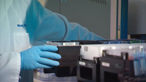
Nodality researchers are utilizing liquid-handling robotics and laboratory automation to improve efficiency and to increase the throughput of single-cell network-profiling assays.
Searching the Secretome
Daniel Chelsky, Ph.D., CSO at Caprion Proteomics, presented his company’s proteomics methodology to biomarker discovery and validation. “We combine a search for biomarkers in biological fluids with discovery in tissues, driven by highly multiplexed assays.”
Dr. Chelsky’s overall developmental strategy is based on MRM, a mass spectrometry assay that can specifically target large numbers of proteins. Promising biomarkers can be assembled by trolling the literature, proteomics assays, and transcript profiling. Through a validation funnel, the candidates are screened and winnowed down to the most promising possibilities.
“From this data, expression in the source tissue predicts expression in blood,” Dr. Chelsky said. “We can also determine the specificity of these proteins by building a ‘Body Atlas’ catalog of shared vs. specific secreted proteins from a wide range of tissues. We believe that this approach, combined with highly multiplexed multiple reaction monitoring, provides a powerful tool for biomarker verification.”
The company is focused on biomarker discovery using label-free, gel-free, quantitative mass spectrometry—a nonhypothesis-driven strategy, or “fishing expedition” in which thousands of proteins are profiled, narrowing down to dozens or hundreds of differentially expressed proteins. Subsequently, these are verified through a targeted approach involving multiplexed, quantitative multiple-reaction monitoring by mass spectrometry. This strategy allows large numbers of samples to be analyzed with a final aim of validation for regulatory compliance.
Caprion has pursued a number of malignant conditions such as lymphoma within the CNS. Diagnosis of this condition is fraught with difficulty, requiring expensive imaging or invasive biopsies that are dangerous and not always possible. As an alternative, cerebrospinal fluid from patients with lymphoma and matched controls was investigated, resulting in some 76 candidate markers. Among these, antithrombin III levels were found to be an accurate predictor of disease progression, providing better sensitivity and specificity than the conventional cytology, according to the company.
Given that secreted proteins are perhaps a thousand times more concentrated in the Golgi apparatus than in the blood, targeting this subcellular organelle and the attendant secretory vesicles to reveal the secretome is another promising strategy to detect markers present in very low concentrations. Caprion staff investigated the presence of biomarker proteins in prostate tumors and their normal counterpart.
Among 60 differentially expressed proteins, they were able to identify seven that were previously identified by other investigators in plasma. Two of these, prostate specific antigen and macrophage migration inhibitory factor, were measured by ELISA in the plasma and were strongly correlated with measurements in the patients’ tumors.
Another series of investigations identified secreted protein indicative of progressive type 2 diabetes. By focusing on those secreted proteins found only in the beta cells, Caprion researchers identified almost 300 proteins specific to the beta cells that are currently being evaluated as candidate biomarkers.
Multibiomarker Profiles
It is well known that between 1950 and the early part of this century, deaths due to cardiovascular insult dropped precipitously while cancer death rates were virtually unaltered. How can we change these dismal statistics?
According to Anna Lokshin, Ph.D., associate professor at the University of Pittsburgh Cancer Institute, “Cancer biomarkers serve many uses ranging from early detection to differential diagnosis to therapeutic monitoring. So because of its lethality in the later stages of the disease, we feel that it is critical to develop new and more effective screening tools.”
Given that the lifetime risk of ovarian cancer is 1.8%, an effective screening strategy must have a sensitivity of at least 80% and a specificity of 99.6% in the early stages of the disease. Dr. Lokshin and her colleagues have designed a serum multimarker panel using a group of well-known biomarkers, which for ovarian cancer yielded a sensitivity of 90% and a specificity of 98%. However, to be effective, higher sensitivity and specificity is required, so there is a need for biomarkers able to recognize preclinical disease at an earlier stage.
“This means we need to examine additional biomarkers, combine different classes of biomarkers, for example, proteins and nucleic acids, or look at biomarkers in other bodily fluids,” Dr. Lokshin concluded. Indeed, for a number of cancers, including ovarian, pancreatic, lung, and breast, marker levels in the urine of healthy individuals compared with that of cancer patients proved to be more accurate in terms of both sensitivity and specificity than in serum.
These studies, while tantalizing, leave open a number of questions for future investigations. This includes a need to verify the performance of expanded sets of serum biomarkers of these cancers while further optimizing the biomarker panels through combinations of serum and urine biomarkers.
Another strategy would be to develop recombinant antibodies that are optimized for urine biomarkers using selective screening of phage-display antibody libraries. Yet another approach would be to examine combinations of urine proteins with DNA isolated from urine or from microRNA.
An alternative task is to initiate prospective collection of matching urine/serum samples in symptomatic patients. Specifically included would be ovarian cancer patients with a pelvic mass, breast cancer patients who are positive on mammogram studies, lung cancers diagnosed through CT scan, and pancreatic cancer diagnosed by MRI. All these materials would be preserved for future validation.
But, perhaps the most challenging need is to convince the principle investigators of large-scale cancer screening trials to collect urine samples from their participants, given the fact that urine has been ignored by cancer researchers over the years as a source of diagnostic information.
Network Analysis of Cancer Mutations
Ali Torkamani, Ph.D., of the Scripps Institute of Genomic Medicine, discussed recent studies on somatic mutations in cancer cells. He stressed that the analysis of the frequency of specific mutations among different tumors has identified a number of mutated genes that contribute to tumor initiation and progression. “Network-based analyses, in which we consider tumor profiles as a system rather than as discrete events, may provide increased power in detecting driver mutations and predictive signatures.”
Dr. Torkamani described this approach in cancer biomarker discovery, both for somatic mutation analysis and the identification of predictive signatures. The group’s protocol applies co-expression, gene ontology, literature, and interaction searches to various cancers. From these studies they derived modules indicating the functional relationships of the mutated genes making up the networks.
“Through this analysis, we identified Wnt/TGF-beta cross-talk, Wnt/VEGF signaling, and MAPK/focal adhesion kinase pathways as targets of rare driver mutations in breast cancer, colorectal cancer, and glioblastoma, respectively,” Dr. Torkamani asserted. “It is our belief that these mutations contribute to a refined shaping or ‘tuning’ of these pathways in such a way as to result in the inhibition of their tumor-suppressive signaling arms, and thereby, conserve or enhance tumor-promoting processes.”
Previous efforts to detect rare driver mutations have focused on known pathways or known direct interactions between mutated genes, resulting in descriptions of tumorigenic processes only in general terms, lacking specificity with respect to the role of these individual mutations in the tumorigenic process.
However, Dr. Torkamani stated that the network reconstruction and gene co-expression module-based approach was based on identifying a larger number of mutated genes than expected by chance. This unbiased approach does not rely on prior knowledge of the biological relationships between genes, but rather attempts to reconstruct sets of coordinately acting genes in order to define, de novo, biological processes affected by cancer mutations.
While Scripps’ approach to identifying cancer biomarkers does not offer immediate clinical applications, it does demonstrate how network-based approaches can be incorporated into a genomic medicine strategy directed toward the understanding of tumor development. These findings can serve a basis for new therapeutic and diagnostic tools.

Scientists at Scripps Institute of Genomic Medicine are using an approach featuring network-based analyses in which they consider tumor profiles as a system rather than as discrete events. They believe this strategy may provide increased power in detecting driver mutations and predictive signatures.
K. John Morrow Jr., Ph.D. ([email protected]), is president of Newport Biotech and a contributing editor for GEN.


