May 15, 2014 (Vol. 34, No. 10)
It sounds like science fiction: fabricating three-dimensional (3D) human tissues using technology very similar to that of a common inkjet printer. Yet bioprinting, as the process is called, has been a reality for over a decade.
The first patent for modified inkjet printing of viable cells was awarded to Thomas Boland, Ph.D., at Clemson University in 2003.
Since that time, the technology has advanced in many areas, uniting such diverse disciplines as engineering, materials science, cell biology, and regenerative medicine. The first company to commercialize bioprinting technology was San Diego, CA–based Organovo, an early-stage regenerative medicine company founded in 2007. Organovo built upon the foundation of cell-printing technology developed by a team of researchers at the University of Missouri-Columbia, led by Gabor Forgacs, Ph.D.
The company’s executive vice-president of commercial operations, Mike Renard, explains that creating functional human tissue through 3D bioprinting must leverage multiple materials and a computer-controlled printed design. “In our case,” says Renard, “the multiple materials are the different cell types that need to be included in the tissue to achieve the specified level of functional performance.”
“The second aspect to be defined is the architecture best suited for the tissue to mature and meet those same specified performance levels,” Renard continues. “It is this combination of composition and printed design that strongly influences the quality and performance of the resultant tissue.”

One of Organovo’s tissue engineers oversees the construction of a vascular tissue construct on the Novogen MMX bioprinter.
Starting Small
The first experiments with printing human cells required an inert material that acted as a scaffold for seeding the cells. This technology formed the basis of the Boland patent.
Early experimenters included Anthony Atala, M.D., director of the Wake Forest Institute of Regenerative Medicine in Winston-Salem, NC. “Our bioprinting research actually began on a desktop inkjet printer that was modified to print 3D structures, recalls Dr. Atala.“Cells were placed in a sterilized ink cartridge. The printers we use today are based on this same concept, but are much more sophisticated and precise.”
Dr. Atala explains that current bioprinting devices must meet exacting standards: “The printers we’ve designed give us the option of using two or more different cell types and placing them exactly where they need to be.” He notes that 3D printers offer considerable flexibility when it comes to the scaffold material—they can accommodate both gel-like or rigid scaffolds, or even dispense with the need for scaffolds altogether.
The latter option is a key element of Organovo’s process. “Our printing technology platform creates 100% fully cellular tissues, free from any scaffolds or added-in materials,” asserts Renard. “We do utilize hydrogels in some circumstances as a mold until we achieve fusion of the printed cellular bioinks. The gel is then removed or dissipates, leaving only a pure tissue as the final construct.” Renard adds that this process “builds a tissue that reproduces a native-like microenvironment.”
Hydrogels are widely used in bioprinting, and extensive research has been devoted to finding the best materials to support the wide variety of tissues being printed. One researcher that has focused on inkjet-printed hydrogels is Paul Calvert, Ph.D., a professor of bioengineering at the University of Massachusetts-Dartmouth.
“My approach to materials development is based on looking for the gaps: what materials don’t exist,” says Dr. Calvert. “If we had strong gels like biological tissues, we should be able to build animal-like machines.”
Dr. Calvert’s laboratory emphasizes flexibility and creativity. “Materials do get developed with a particular goal in mind, but that is almost never what they are used for in the end,” Dr. Calvert observes. “Kevlar was developed to be tire cord. Polyethylene was intended for high-tech insulation. Polylactic acid is getting popular for 3D printing but was originally intended for biodegradable packing.”

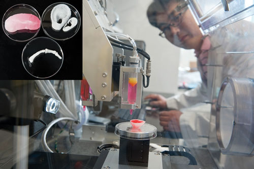
Hyun-Wook Kang, Ph.D., an instructor at the Institute for Regenerative Medicine at Wake Forest Baptist Medical Center, monitors the progress of a bioprinted kidney structure. The inset shows a kidney prototype as well as ear and finger bone scaffolds.
Bioprinting Tissues
While it’s tempting to fantasize about the ultimate goal of the technology—bioprinting replacement tissues and organs—even printing living cells can be a delicate business.
“Challenges include ensuring that the printing process does not alter the cells,” says Dr. Altala. In addition, the printed structure and its cells “must remain viable until it can be implanted in the body.” These challenges are magnified when working on a larger scale, Renard explains: “A second challenge related to building larger mass/volume tissues is the need to include a vascular or capillary network to feed the tissue without cell death in the interior.”
One of the immediate benefits of bioprinting lies in helping researchers improve the drug discovery process. Dr. Calvert cites a short-term goal to bioprint a small section of viable liver tissue that can be used to test for patient-specific drug responses. He sees the potential for bioprinting complete organs down the road, but notes that “lab samples for testing, diagnosis, and customized production of active proteins are all much closer.”
Dr. Atala also sees this as an opportunity. “With collaborators at other institutions, we are working to print miniature hearts, lungs, blood vessels, and livers onto ‘chips’ that will be connected with a blood substitute,” he reveals. “Called a ‘body on a chip,’ the system has the potential to speed up the development of new drugs because it could potentially replace testing in animals, which can be slow, expensive, and not always accurate.”
Bioprinting Tissues
Toward Replacement Organs
Other researchers are embracing the challenges of bioprinting complete, fully functional organs. One of these researchers is Stuart Williams, Ph.D., division chief, Bioficial Heart, Cardiovascular Innovation Institute in Louisville, KY.
For Dr. Williams, the research is personal—his family history of cardiovascular disease inspired his decision to focus on the field as an opportunity to apply bioprinting technology. He explains, “I have always been interested in the microcirculation, and I saw bioprinting as a way of creating microcirculations with defined structures. And finally, I see bioprinting as a way of creating totally biologic organs—what I call bioficial organs.”
Dr. Williams envisions a future where the bioficial heart will be used in therapy to treat heart disease, but meanwhile he sees printing a heart patch that provides blood flow to an ischemic region of the heart as an incremental achievement: “Parts of hearts may also be critical toward the treatment of pediatric heart disease—treating openings between chambers in the hearts of children (septal defects).” Dr. Williams notes that pieces of the child’s own tissue could be combined with bioprinted tissue to provide a form that matches the defect.
When it comes to prosthetic implants, Dr. Williams’ pioneering research with microvascular endothelial cells provides the key to ensuring that the implants are successful. “Microvascular endothelial cells are the most critical part of implants,” emphasizes Dr Williams. “Without the microcirculation, implants will not survive.”
Other tissues are being targeted, under Dr. Atala’s oversight, for clinical applications. “For the Armed Forces Institute of Regenerative Medicine [a $75 million federally funded effort to apply regenerative medicine to battlefield injuries], we are working to print muscle, bone, and cartilage tissue that could be used in reconstructive surgeries,” relates Dr. Atala. “We are also working to print skin cells onto burn wounds as an alternative to skin grafting.”
Future Directions
Given the rapid pace of advances, are we likely to see bioprinted organ banks ready for transplant any time soon?
Renard suggests a disciplined approach for Organovo: “Much more research and progress is needed before the idea of replacement organs on demand could be a reality. We envision a progressive advance from the smaller and simpler designs, such as tubes and patches that repair and regenerate, to the larger and more complex that replace function.”
Dr. Williams is optimistic when it comes to his vision for treating cardiovascular disease. “The hope is that we can develop a universal cell that can be used to bioprint organs and then bank these organs for use in all patients,” he explains, adding that the idea of growing organs outside the body was first described by Charles Lindbergh—the aviator—a century ago.
Dr. Williams describes the application of bioprinting to prosthetics as a stepwise process: “At first, the printed devices will not contain cells—we already see this technology moving into medical and dental practice. The next step will be 3D printed devices with cells incorporated—something we call hybrid devices. Watch for this to appear in orthopedic and dental implants. Then, parts of organs created using completely biologic components. And finally, completely bioficial organs bioassembled in the operating room.”
Dr. Atala cites the challenges associated with supplying oxygen to bioprinted organs while they integrate into the body. “One possibility is to print small channels into the structures that can be populated with blood vessel cells,” he point out. “Another option might be to print oxygen-producing materials into the scaffolds.”
He reflects that the ability to print organs on demand would address many of the shortcomings of the current organ banking and transplant system, including the lack of donor organs and the need for powerful immunosuppressant drugs that have side effects. “Whether tissue engineering can replace organ banking, I cannot predict,” shares Dr. Atala, “but I think it certainly has the potential to one day augment it.”
Want more on bioprinting? Be sure to check our GEN Exclusive “Patentability of 3D-Printed Organs“.


