August 1, 2012 (Vol. 32, No. 14)
Molecular imaging plays a key role in pharmacology evaluation and is expected to grow significantly in the future. Even though preclinical imaging has proven its worth, it is still a relatively new technology. Consistent paradigms have yet to be developed and applied to drug discovery and development.
As instrumentation and software continue to improve, new tracers develop, and multi-modality approaches refine, proof-of-concept in the preclinical setting will likely translate into valuable clinical diagnostic tools.
Novel advances in molecular imaging for preclinical applications were discussed at the recent “WorldPharma Congress” conference.
Quantitative whole-body autoradiography (QWBA) is widely used by the majority of the pharmaceutical industry to provide quantitative tissue distribution data as part of a preclinical ADME program.
Conventionally, QWBA is used to support clinical study safety by ensuring that drug and associated radioactivity exposures, based on a predicted dose level, are appropriate for human testing.
“Being able to quantitate how much drug or drug equivalent you have, based on a radioactive tag in a particular tissue three-cell-layers thick, has increased the drug safety and tissue distribution aspect 10-fold,” commented Stefan Linehan, manager, preclinical services, XenoBiotic Laboratories, and president, Society of Whole Body Autoradiography.
If the radioactive concentration is low or at a similar concentration in adjacent tissues, as displayed on a standard gray-scale autoradioluminogram, the mapping out and outlining of the regions of interest can be best-guess determined, if at all. Linehan addresses these resolution issues with a technology he terms cryo-imaging and quantitative autoradiography (CIQA).
CIQA allows for a series of optical images, captured every 25 µm, throughout the whole body of a frozen rodent carcass embedded in a solid block of carboxymethylcellulose. Periodic consecutive sections are acquired and processed to generate informative images produced by autoradiography, histology, fluorescence, and immunohistochemistry.
Using a customized software program, the images are registered and reconstructed in 3D, allowing entire organs to be viewed in high resolution, 25–50 µm, with all the informative sections interlaced.
In addition, the long half-life of the radioisotopes used, such as 14C, allows for pharmacokinetic and pharmacodynamic models within different tissues over an extended timeframe.
“CIQA is meant to be a complementary technique, to help add to the knowledge gained from other imaging modalities. The recent advancements can be used to provide useful information in many other fields such as toxicology, pharmacology, neurology, and oncology,” concluded Linehan.
Maximizing Information Extraction
Extracting as much encoded information as possible from an image, including deriving kinetic parameters, is crucial in molecular image analysis.
In pharmacokinetic groups, a common way to analyze data is measurements of signal changes over some period of time, the area under the curve (AUC). However, that analysis does not capture the kinetics.
“We use DCE-MRI to investigate anti-angiogenic and antivascular therapies for tumors because it allows us to measure tissue perfusion and permeability,” discussed Matt Silva, Ph.D., director, research imaging sciences, Amgen.
“The problem with dynamic-measuring methods is that in order to fit to a compartmental model, we need to know the input function to the system, the injected contrast agent, and how it transverses through the body. There is no easy way to do that.”
Current input method estimations—location of a vessel, population-based averages, or the use of reference regions—have flaws. They do not capture the kinetics, and this is why researchers using DCE-MRI extended the analysis beyond AUC into the Ktrans value.
“Utilizing the target tissue as an estimate for the input function for modeling may be a more sensitive way to detect changes and has the added advantage of enhancing the performance of the software. This methodology is not specific to DCE-MRI and can be used in other modalities, like FDG PET. You can measure AUC, but if you want to extract the metabolic rate of glucose you need to do compartmental analysis.
“There are additional challenges in imaging analysis. We spend weeks doing whole-body analyses. Half our work is data collection and half analysis. Looking at specific regions, or organs, during data analysis can be very time consuming,” continued Dr. Silva.
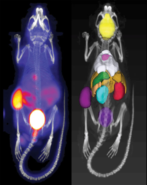
“So we initiated a digital mouse atlas project. The concept is to be able to quickly screen with the digital atlas and rank order on semi-automated analyses. Then our scientists would step in and evaluate the most critical datasets.”
In collaboration with inviCRO, the digital mouse atlas project is still in early phases, but the proof-of-concept is promising.

PET imaging probes distribute to tissues proportional to perfusion and permeability as well as target concentration and activity. The image on the left shows the thymidine analog 18F-FLT, which distributes not only to tumor (due to high rate of cellular proliferation) but also to GI and bladder (due to clearance). The analysis of individual regions (e.g., organs and lesions) is time-consuming labor, which may be accelerated with computer models of mouse anatomy, shown on the right. [Amgen]
Quantifiable Lung Imaging
Diffuse lung diseases, such as emphysema and pulmonary fibrosis, are very difficult to quantify. Historically, histological sections were used, but unless the entire lung is sliced, quantification can be hampered.
“Improvements in CT devices allow imaging of soft tissues, like the lungs, with fairly good resolution,” explained Lawrence de Garavilla, Ph.D., scientific director and fellow, imaging and PK/PD, immunology research, Janssen Research and Development. “With our micro-CT unit we can get in vivo resolution of 45–50 µm and in ex vivo studies, using a contrast agent, 5 µm. This allows us to look at alveoli, the terminal structures in the lung where gas exchange occurs.
“CT imaging allows us to image the whole lung, create a 3D rendering, and quantify the amount of fibrosis. It is a big change; we now have an objective quantifiable technique. Instead of having a pathologist sit at a microscope and subjectively analyze 50–100 lung slides, the resolution of our assay and the robustness of this imaging technique allow determination of, and differentiation between, 5 and 10% degree of fibrosis.”
Measuring efficacy during pulmonary drug clinical trials is very difficult. Functional tests, such as spirometry, are used but are crude measures of airway inflammation, fibrosis, and emphysema. Imaging techniques could prove to be a valuable clinical tool to quantify the degree of lung disease and improvement with novel therapeutics.
Looking forward, CT imaging may also be applicable as a noninvasive clinical diagnostic tool, especially for airway inflammation. Currently, bronchoscopies are performed but only on a limited basis since they are invasive and provide incomplete diagnostic information.
Dr. de Garavilla envisions using lung imaging to track the number and different types of inflammatory cells, to differentiate which cells are going into the lungs and the therapeutics’ effects on the various cell types.

Emphysematous mouse lung: histological section, micro-CT gray-scale image, and gray-scale image with green highlights of emphysematous alveoli. Images convey the transition of classical methods to evaluate lung disease, histology, to CT imaging and image-analysis techniques. [Janssen Research and Development]
Tracking Molecules and Diseases
PET tracers can be target- or disease-based. “Target-based PET tracers are used to determine that you have the desired characteristics of the pharmacological molecule. This is an essential step in developing a molecule and being able to take it into proof-of-mechanism or proof-of-concept studies,” said Aidan Power, M.D., vp and head, pharmatherapeutics, precision medicine, Pfizer Worldwide Research and Development.
“Let’s say a compound must bind to 80% of a particular receptor in the brain to be efficacious. We need to use a tracer so we can understand the percent of occupancy of the receptor. That is why there is a specific need to develop PET tracers for specific receptors.”
Required properties for the target-based tracers depend on where they will go in the body. PET tracers that need to go into the brain must have a molecular weight less than 600 and be moderately lipophilic. Target-based tracers must be highly specific, have high affinity, accumulate in target-rich tissues, and clear rapidly from others.
“There is also a lot of interest in developing PET tracers for disease processes. The amyloid-plaque tracers are disease-based, not specific to a particular drug, and may well be used for diagnosis in the future. At least you have it reasonably narrowed down that you have a strong clinical suspicion that someone has a disease. It is possible you might use them to evaluate how well someone is responding to treatment,” continued Dr. Power.
“We are going to continue to see a lot of development in tracers to help us diagnose and understand disease. These are significant advances that will require a lot of science. It is not so much about the specifics of the technology as much as about how we will be able to systematically apply it.”
The Cheap Man’s PET
Cerenkov radiation is the familiar blue glow one sees coming from a pool of water in a nuclear reactor.
In 2009, using a modern ultra-cooled CCD technology, the first Cerenkov emission from a subcutaneous tumor after in vivo administration of the commonly used PET tracer 18F-FDG was recorded. Data showed accumulation of the tracer in tumor-bearing animals by imaging its optical signal, rather than its nuclear decay signature.
While bioluminescence is a chemical reaction that creates light, CR is light produced at the quantum level.
“If we look at the nuclear decay of beta+ or beta- events, one of the things released is a charged particle. As the charged particle interacts with the local medium it polarizes the surrounding area, and after it passes, the area relaxes back to its normal state. During this transition CR is produced. Light is the resulting output,” explained Robbie Robertson, scientist I, biomedical imaging group, Millennium: The Takeda Oncology Company.
Unlike PET, which measures an annihilation event that creates gamma rays, CR is produced along the entire path length of the charged particle.
“Our throughput capacity of Cerenkov luminescence imaging (CLI) is approximately 10-fold greater than traditional PET imaging. Plus, within a few hours of training, anyone can run a CCD system.”
“One of CLI’s greatest advantages lies in its ability to allow a company to quickly screen through a large amount of radiolabeled compounds in a shorter timeframe than is currently achievable with preclinical PET imaging systems. This frees up time on PET scanners and allows them to be used to produce the required higher quality data for future transition of a drug from the preclinical to the clinical setting,” concluded Robertson.
Affectionately referred to as “The Cheap Man’s PET”, the number of articles referencing CLI since 2009 illustrates the modality’s acceptance.

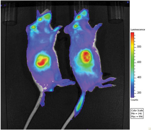
A Cerenkov luminescence imaging (CLI) image is shown of control animals at 29 days, with a subcutaneous tumor (OCI-LY10 lymphoma cell line) on the right flank. [Millennium: The Takeda Oncology Company]


