April 15, 2009 (Vol. 29, No. 8)
Improving Image Acquisition, Reconstruction, Visualization, and Analysis
Preclinical imaging is rapidly becoming a standard technology for drug discovery and development. This adoption has been driven by the increasing pressure on pharmaceutical companies to identify ways to reduce costs, increase safety and efficacy, and minimize side effects.
Preclinical imaging technologies, when used appropriately in the right stage of the drug discovery process, can contribute significantly to efficiency gains by identifying failures early, providing safety and efficacy data based upon in vivo results, gathering insight into long-term effects using small animals with much shorter life spans, and minimizing the number of animals used by virtue of being able to perform longitudinal studies.
Although a number of imaging modalities are commonly used, technologies that utilize radioactive tracers such as positron emission tomography (PET) and single photon emission computed tomography (SPECT) have found widespread acceptance due to high sensitivity, excellent spatial resolution, and the ability to perform dynamic studies. Hybrid technologies that incorporate computed tomography (CT) have also become a standard component of preclinical imaging studies.
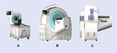
The Inveon® family of imaging systems from Siemens encompasses PET, SPECT, and CT technologies (Figure 1). The Inveon preclinical imaging systems also include Inveon Acquisition Workplace (IAW) and Inveon Research Workplace (IRW) software solutions.

Figure 1. Inveon family of preclinical imaging scanners
Preclinical Imaging Workflow
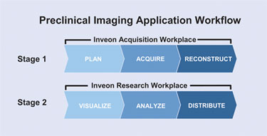
Recent imaging advances have helped to speed up both basic preclinical research and drug discovery research. As for any transformational technology, however, it is important to incorporate this technology into the existing workflow with minimal disruption. An understanding of the preclinical imaging application workflow is critical to this process. This workflow can be broken down into two stages (Figure 2). Stage 1 can be further divided into three distinct steps, primarily focusing on image acquisition and reconstruction of acquired data.
• Plan—The details of the image acquisition and reconstruction protocols are set up depending on the technology that is used, which, in turn, dictates the experimental workflow.
• Acquire—The experimental subject is placed on the scanner bed and image data is acquired. In a PET or SPECT study, the gamma photons emanating from the radiotracer injected into the subject are detected along with positional information. In the case of a CT study, the x-ray source emits a beam that transverses the subject. The attenuated photons are detected by the detector and the intensity and positional information are stored.
• Reconstruct—The stored data is then reconstructed to form a 3-D image volume, with several different choices of reconstruction algorithms. Reconstruction methods vary depending on the specifics of the data that were acquired.
Stage 2 can also be divided into three distinct steps, primarily dealing with visualization of images and quantitative analysis of regions of interest.
• Visualize—Reconstructed images from a single subject acquired over several days or multiple experimental subjects may be viewed simultaneously to identify any changes and regions that require further quantitative analysis. Data that was processed into dynamic and gated or gated-dynamic image volumes can also be viewed.
•Analyze—Regions of interest that are identified from the previous step can be subjected to quantitative statistical analysis including calculating the mean activity value, volume, and surface area for a region. Additionally, time-activity curves can be generated to track the pharmacodynamics of radiotracers and to perform pharmacokinetic modeling as required.
• Distribute—Finally, the data can be exported to several formats for archiving and future analysis. It is also possible to create movie files from dynamic and gated image data, generate graphs, and capture specific images for distribution or storage.
A typical preclinical imaging experiment in drug discovery applications involves multiple studies in parallel utilizing several experimental subjects. The compounded effect of the time invested could easily impact the overall efficiency gains and cost advantages despite the overarching benefits of this technology.

Figure 2. Typical preclinical imaging application workflow
Inveon-Workplace
Siemens Preclinical Solutions recently launched Inveon•Workplace, a preclinical imaging solution with features that directly enhance workflow efficiency. Inveon• Workplace includes the latest versions of IAW and IRW with significant enhancements to image acquisition, reconstruction, visualization, and analysis functionalities.
Enhancements to the image acquisition speed in the new version of IAW were accomplished for all three modalities—PET, SPECT, and CT—which directly translates to less time required for this step of the workflow. In the case of PET, changes were implemented in the way data is transferred from the scanner to the embedded PC, which significantly reduce the loss of data packets.
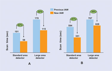
The option of multipinhole collimators in the SPECT system increases the sensitivity of the system, which allows for the acquisition of the same number of statistical data points in a shorter amount of time. Finally, the speed of anatomical resolution CT scan speed was increased by almost a factor of two as compared to the previous version of IAW (Figure 3A, standard area detector). The scan speed for medium-resolution CT scans was also improved in the new version of IAW (Figure 3B, standard area detector).
Enhancements to image reconstruction techniques in the new version of IAW for PET, SPECT, and CT modalities results in a higher quality of reconstructed data, and thereby less time spent in assessing the image quality or repeating studies to obtain high-quality image data. Use of component-based normalization for PET scans delivers higher quality data with higher statistics in a much shorter scan time. In addition, the CT-based attenuation correction option minimizes noise in the PET image and improves quality. CT-based attenuation correction is now available as an automated workflow.
The quality of the SPECT image data is enhanced by virtue of helical scanning capability, which leads to fewer artifacts in the reconstructed images. The axial field of view is also increased by adding more helical turns; this allows whole-body scans in a single experiment. The overall stability of high-resolution CT image reconstructions has been significantly enhanced with new version of COBRA real-time reconstruction engine.
A number of enhancements were implemented in the loading and handling of PET and CT data, which reduces the overall time invested in image visualization and analysis steps. The latest version of IRW operates on a 64-bit processor so the software can make optimal use of the 16 gb RAM in the workstation.
The end result is that data volumes as large as 8 gb can be handled with ease by the new version of IRW.
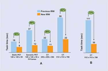
Enhancements to the data-handling feature of IRW enables loading of static and dynamic PET and CT images at least two times faster than previous versions (Figure 4A). With the new version of IRW, images acquired by Inveon imaging systems can be launched directly without the need for DICOM conversion, resulting in a reduction in image load time (Figure 4B).
Inveon•Workplace enhances the overall preclinical workflow by maximizing efficiency. Improvements in speed, quality, and performance facilitated by IAW and IRW will speed up preclinical imaging studies.

Figure 3. Enhanced CT scan speeds with new version of IAW

Figure 4. Enchanced speed of PET and CT data loads and direct CT data launch with new version of IRW
Santosh S. Arcot, Ph.D. ([email protected]), is marketing manager, Preclinical Solutions, Siemens Medical Solutions. Web: www.siemens.com.


