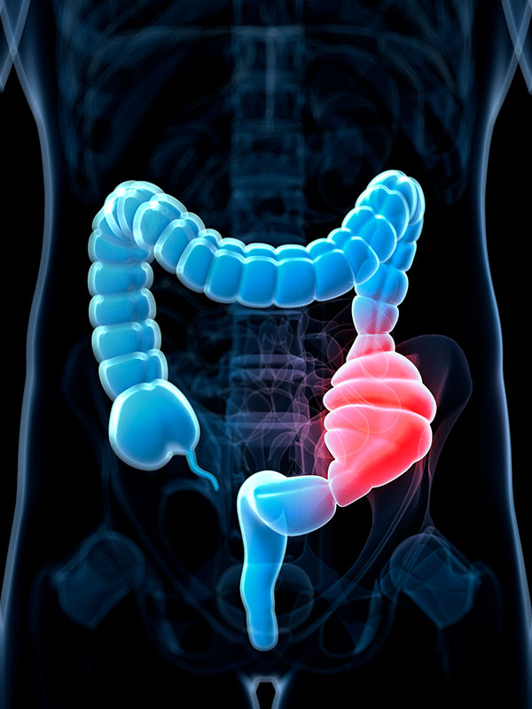
It is fairly common to think of constipation in a humorous light. However, anyone who has suffered from the debilitating condition can attest that it is far from a laughing matter. With a sizable percentage of the population increasing in age and opioid abuse reaching epidemic proportions, investigators are looking for new tools to help patients ease their gastrointestinal suffering. Now, investigators from Thomas Jefferson University (TJU) have just released data of a new technique—called magnetofection—that incorporates micro-metal beads coated with small RNA fragments (microRNAs, or miRNAs) injected at specific regions of the colon and held in place with a powerful magnet.
The findings from the new study were published recently in the American Journal of Physiology—Gastrointestinal and Liver Physiology in an article entitled “In Vivo Magnetofection: A Novel Approach for Targeted Topical Delivery of Nucleic Acids for Rectoanal Motility Disorders.”
Both constipation and incontinence, although multifactorial, have also been associated with the problems related to the regulation of a ring of smooth muscle around the anal opening, termed the internal anal sphincter. Unlike the way we can clench muscles just by thinking about them, smooth muscle is a type of muscle that we can't control with our thoughts. Using this new technique to deliver gene-therapy-like intervention directly where it's needed, the TJU researchers successfully increased or decreased the muscle tone of the anal sphincter in appropriate animal models. These studies could have implications in treating constipation or incontinence.
“…we developed a novel approach of in vivo magnetofection for localized delivery of nucleic acids such as micro-RNA-139-5p (miR-139-5p; which is known to target Rho kinase2) to the circular smooth muscle layer of the internal anal sphincter (IAS),” the authors wrote. “The IAS tone is known to play a major role in the rectoanal continence via activation of RhoA-associated kinase (RhoA/ROCK2). These studies established an optimized protocol for efficient gene delivery using an assembly of equal volumes of in vivo PolyMag and miR139-5p or anti-miR-139-5p (100 nM each) injected in the circular smooth muscle layer in the pinpointed areas of the rat perianal region and then incubated for 20 min under magnetic field.”
Rather than simply inject the agent, where it could have spread through the tissue to affect other regions, the investigators kept the miRNAs localized to the ring of muscle. The research team first injected the miRNAs mixed with micro-metal beads, and then kept them localized to the sphincter with a magnet. This innovative yet simple approach was more effective at keeping the miRNAs in the right place than injecting the miRNAs alone.
“Magnetofection efficiency was confirmed and analyzed by confocal microscopy of FITC [fluorescein isothiocyanate]-tagged siRNA [small interfering RNA],” the authors noted. “Using physiological and biochemical approaches, we investigated the effects of miR-139-5p and anti-miR-139-5p on basal intraluminal IAS pressure (IASP), fecal pellet count, IAS tone, agonist-induced contraction, contraction-relaxation kinetics, and RhoA/ROCK2 signaling.”
Because sphincters also help keep food in the stomach from rising up the esophagus, using a similar approach has additional implications in the lower esophageal sphincter where its localized stimulation could help prevent acid reflux in difficult-to-treat cases. First, however, the researchers will have to demonstrate whether the same principles hold true in humans, without side effects.
“Collectively, these data demonstrate that magnetofection is a promising novel method of in vivo gene delivery and of nucleotides to the internal anal sphincter for the site-directed and targeted therapy for rectoanal motility disorders,” the authors concluded.


