
May 15, 2010 (Vol. 30, No. 10)
Technological Advances Continue to Increase the Versatility of This Reliable Lab Workhorse
The “Ion Chemistry and Mass Spectrometry Conference” held at Lake Arrowhead, CA, earlier this year, covered many advances in mass spectrometry that have allowed unprecedented analysis of molecular events occurring in normal and pathological biomolecular systems.
Among the enabling technologies highlighted at the meeting was work done by Joseph Loo, Ph.D., professor of chemistry and biochemistry of the University of California, Los Angeles. “We have developed electrospray ionization mass spectrometry methods with improved sensitivity for detecting and measuring protein noncovalent complexes,” he said.
His group is presently also concerned with obtaining structurally relevant information on protein-ligand interactions. In addition, the team, which previously reported a mass spec-based method for localizing ligand binding sites, has now extended this work to address nucleotide binding, specifically aiming at the site of ATP-binding to adenylate kinase.
Mass spectrometry is an outstanding tool for investigating molecular interactions—it is sensitive, specific, and fast, particularly when combined with electrospray ionization, which ionizes macromolecules without disrupting weak, noncovalent interactions. The extremely precise molecular mass measurement provides direct evidence for protein-ligand or nucleotide-ligand associations. Indeed, the interactions may be retained during the transition from liquid to gas phase, thus permitting the size of the complex and the binding stoichiometry to be accurately assessed.
Classically, mass spec analysis uses a “bottom-up” strategy, in which molecules are fragmented with proteases, then their molecule weights are assessed, and an overall picture of the original protein is assembled. However, Dr. Loo and his colleagues are exploiting a “top-down” approach to ligand-protein dynamics in which intact complexes are introduced into the mass spectrometer, so information describing the native complex is retained.
Even molecules that bind relatively weakly to one another can maintain their association in the gas phase, the relationships being fixed by the solution-phase positioning of the component molecules. Dr. Loo has taken advantage of the stability of electrostatic interactions to determine the adenosine 5´ triphosphate binding site of adenylate kinase.
“We have also discovered new reagents for increasing protein charge for electrospray ionization mass spectrometry that markedly improve the efficiency of tandem mass spectrometry of proteins.”
A number of neurological disorders are the result of abnormal protein aggregation, including Alzheimer, Parkinson, Creutzfeld-Jacob, and Huntington diseases. Normal protein components are modified in ways that force their aggregation, blocking normal neuronal impulse transmission. An understanding of this process, obtained through the use of electrospray ionization mass spectrometry, could contribute to the design of therapeutic molecules for these conditions.
Dr. Loo has used data from his top-down mass spec strategy to probe the sites of compounds binding to amyloid b protein. Amyloid b self-aggregates into a variety of neurotoxic structures from fibrils to oligomers, which are the more neurotoxic of the two. A potential anti-Alzheimer therapy could be to develop low molecular weight compounds that block the formation of these toxic aggregates.
“In the development of novel inhibitors, localization of their sites of interaction on the target of interest is a critical first step in the drug discovery process. Our mass-spectrometry studies can augment the existing body of knowledge for a given biochemical complex, and they can provide key preliminary insight on the structure of supramolecular assemblies, especially for complexes not amenable to x-ray crystallography or NMR.”
Yellow Fever Vector
Vicki Wysocki, Ph.D., professor of chemistry at the University of Arizona, Tucson, provided a proteomics analysis of iron-related ovarian proteins from Aedes aegypti, the mosquito responsible for transmitting yellow fever and dengue fever. As the mosquito requires an iron-containing blood meal to carry out oogenesis, Dr. Wysocki and her colleagues reasoned that the iron transport proteins could be a target for vector intervention. Accordingly, they searched for iron-regulated and iron-transport proteins in the ovaries of mosquitoes raised on different diets.
This was another example of the bottoms-up approach, in which the ovaries of the insects were dissected and separated on one-dimensional gels. The bands were excised, digested with proteases, and subjected to liquid chromatography followed by mass spec analysis. The identified proteins were then surveyed on the protein database.
A large number of proteins was identified. Ferritin was one of the proteins found in the ovaries of the mosquitoes raised on the iron-containing diet, suggesting that it is upregulated in the presence of hemoglobin in artificial diets. The presence of the enzyme aconitase, known to require iron for activity in other organisms, was also detected.
“We found that proteins expressed in the ovaries of mosquitoes varied significantly depending on the diets these mosquitoes received,” explained graduate student Michelle Huang, who was involved with the study. “The proteins expressed included essential proteins performing developmental, structural, and ribosomal functions.”
Interestingly, iron-transport proteins did not pass the filtering criteria that the team had designed as part of the experimental protocol. “Some of these proteins could represent targets for population control. We are currently expanding on these experiments for more confident identification.”
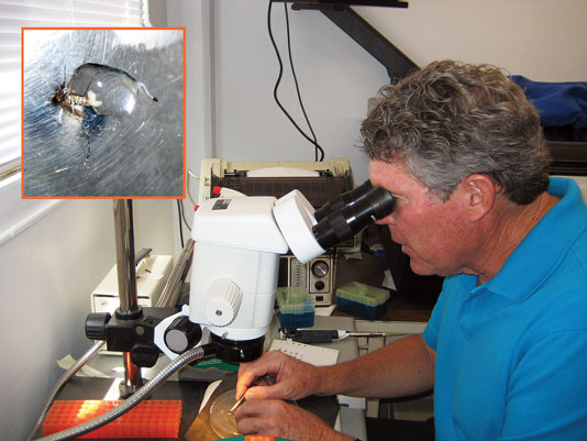
Researchers at the University of Arizona are studying Aedes aegypti, the mosquito responsible for transmitting yellow fever and dengue fever. The inset shows a dissected mosquito in a droplet of protease inhibitor cocktail, showing the mid-gut as well as ovaries removed from the abdomen.
Blocking The Memory Thief
Michael Bowers, Ph.D., professor in the department of chemistry and biochemistry at University of California, Santa Barbara, reviewed the structure and assembly of prion protein fragment PrP106-126. All prion-based disorders are characterized by the accumulation of an abnormally folded form of the prion protein as β-sheet-rich amyloid fibrils in the brain. This pile-up of aggregates in neuronal cells is believed to be responsible for the memory loss that is the hallmark of these devastating conditions.
The early events in the formation of these β-sheets are not well understood. The development of effective vaccines or molecules that could block the aggregation process will require the intimate knowledge of these changes. Dr. Bowers and his team employed ion mobility spectrometry combined with mass spec to delineate one of the key domains involved in amyloid formation.
The team’s observations on monomeric and oligomeric structures of aggregating and nonaggregating forms of the critical region were crunched using replica exchange molecular dynamics. This computer simulation model analyzes the interactions of atoms and molecules over time by approximations from physics, giving a view of the kinetic motion of the particles. The Bowers analysis also employed replica exchange, a Monte Carlo simulation approach that runs parallel replicas of the simulation, selecting the most probable outcomes.
Based on the data collected and the analyses conducted, the team concluded that ordered β-hairpin monomers form the nucleus of the aggregates rather than the converse hypothesis, which states that the process is driven by disordered monomers. This ordered collection of β-structured aggregates then progress to the fibrils responsible for the actual disease. Since observations of the behavior of different forms of the peptide argue for intermolecular rather than intramolecular initiation of oligomer formation, the understanding of the mechanism of the β-sheet-rich amyloid fibrils is moved to a new level.
The analysis performed by the Bowers lab has a broad range of applications outside the field of Alzheimer investigation. A thorough understanding of the dynamics of chemical interactions is a powerful informative tool that will aid in rational drug design and could serve as a means of modeling effective antigens for vaccines.
![]()
Shimadzu Scientific Instruments’ LCMS ion trap time-of-flight (LCMS-IT-TOF) mass spectrometer
Probing Peptide Properties
Mass spec is also proving to be a highly sensitive tool for delineating the effects of charge distribution on peptide structure, which, in turn, affects the behavior of signaling peptides in regulating gene expression. Jianhua Ren, Ph.D., associate professor at the University of the Pacific, and her colleagues presented research on the acidities of short peptides and how this property affects their propensity to undergo conformational changes.
Dr. Ren’s group studies thioredoxins, ubiquitous proteins found throughout living organisms. These enzymes perform as antioxidants by cysteine thiol-disulfide exchange, catalyzing protein reduction. Cysteine residues are found in the active site of the thioredoxin molecule in a CXXC configuration, and they are essential to its antioxidant performance.
Dr. Ren and her colleagues are exploring the helix macrodipolar effects on the gas-phase acidity of peptides. The protein alpha helix has an intrinsic macrodipole moment running from the N-terminus to the C-terminus. In a solution with relatively low pH, the N-terminal cysteine will be ionized and the C-terminal cysteine will not. The ionized cysteine facilitates the thiol-disulfide exchange reaction.
By generating a family of related peptides, Dr. Ren and her team were able to investigate the effect of various amino acid substitutions on the gas-phase acidity of the peptides. “We found that N-terminal cystine peptides are more acidic than the corresponding C-terminal ones, and that longer peptides are more acidic than the corresponding shorter ones,” Dr. Ren explained. “Finally, our computational studies showed that the ionic N-terminal cystine peptides prefer helical conformations, while the ionic C-terminal cysteine analogues are mainly random coils.”
A number of pressing questions in rational drug development can be explored most expediently using mass spec and its many permutations. The discovery process requires refined molecular analysis that enables the modeling of compounds that precisely target the source of the disease process. This may prove to be the best route to achieving a strong pipeline of innovative drugs.

Hybrid instruments combining ultraperformance LC (UPLC) with mass spectrometers play an important role in all aspects of early- and late-stage drug discovery. As the footprint of these instruments continues to decrease in size, the feature-set becomes ever more sophisticated, allowing scientists to derive more information from a single analysis, according to Waters.
K. John Morrow Jr., Ph.D. ([email protected]), is president of Newport Biotech and a contributing editor for GEN. Web: www.newportbiotech.com.

