November 1, 2013 (Vol. 33, No. 19)
It was 30 years ago when Kary Mullis conceived the chain reaction that looked possible by adding polymerase to a DNA molecule in the presence of short oligonucleotide primers and nucleotide triphosphates.
Cycling the temperature to sequentially denature the double-stranded DNA template, anneal primers to the template, and extend the primers to create a replica produced millions of copies. Kary initially had difficulty attracting support from his colleagues and bosses then at Cetus, and he struggled to publish his ideas.
But PCR eventually developed into arguably the most important method in biotech, and Kary was awarded the 1993 Nobel Prize in Chemistry.
When Russell Higuchi’s group accidently ran PCR in the presence of ethidium bromide, which in those days was a common DNA stain for post-PCR electrophoretic analysis, they thought the process would be inhibited due to ethidium being known to be a potent mutagen that binds DNA strongly.
In fact, the reaction was not totally inhibited. The sample began to fluoresce, and the fluorescence increased as the reaction progressed. The Higuchi team realized it should be possible to monitor the amplification in real time through the dye’s fluorescence.
By monitoring the number of amplification cycles needed to produce the amount of product that generates certain threshold fluorescence signals, PCR became quantitative. Initially, the technique was called kinetic PCR, but with time it became known as real-time quantitative PCR, or qPCR for short.
The ethidium bromide was soon replaced with asymmetric cyanine dyes, such as SYBR Green I, that are less inhibitory. Dyes are sequence nonspecific reporters and bind to all products formed, including any aberrant primer-dimer products. This makes dyes less suitable for multiplex analyses and for clinical diagnostics where false positives are of major concern.
PCR products can be distinguished using sequence-specific reporters. The hydrolysis probe, also known as Taqman®, was the first probe invented. It is an oligonucleotide labeled with a fluorescent dye and a quencher that are in proximity in the intact probe such that the fluorescence is quenched. Upon primer extension, the Taq polymerase 5’→3′ exonuclease activity degrades the probe, which releases the dye that starts to fluoresce.
Single-Base Detection
PCR is highly specific and can distinguish between templates that differ in a single base only. The variable position can be interrogated using primers, probes, or post-PCR analysis of the product by high-resolution melting. What’s especially challenging is to study rare sequence variations, such as somatic mutations.
Specificity to detect one mutated sequence against a background of thousands of wild-type sequences is achieved by techniques such as castPCR, myT primers, RNaseH-dependent PCR, and SuperSelective Nucleic Acid Amplification Primers.
Genetic variation does not explain all observed hereditary phenomena; other mechanisms that do not directly depend on DNA sequences contribute. These are collectively referred to as epigenetic. They regulate cell differentiation and include DNA methylation and histone modifications.
PCR can be used to interrogate DNA methylation status either by treating with bisulfite, which converts cytosine to uracil, while 5-methyl cytosine is resistant, or by using methylation-sensitive restriction enzymes. Interaction between histones and DNA can be studied using chromatin immunoprecipitation.
Already 15 years before PCR, Howard Temin and David Baltimore independently discovered the enzyme reverse transcriptase (RT) which is a RNA-dependent DNA polymerase that uses an RNA template to produce a single-stranded DNA transcript known as complementary DNA, which then can be quantified by qPCR.
The yield of RT activity depends on template, priming strategy, and reaction conditions, and ranges between 0.5 and 80%. Performed at optimized conditions, however, RT is almost as reproducible as qPCR, allowing highly sensitive and accurate quantification of messenger RNAs and other long RNA species.
MicroRNAs are small, noncoding, single-stranded RNAs involved in posttranscriptional regulation that were discovered in 1993 by Victor Ambrose. Because they are short (22–24 bases), microRNAs are hard to quantify. Current techniques either use sequence-specific tailed RT primers to produce cDNA that is longer than the template microRNA, use thermodynamically stabilized LNA primers, or extend the microRNA first by polyadenylation or ligation.
Proteins are traditionally detected by antibodies. In 1992 Charles Cantor tagged the detection antibody in an ELISA setup with an oligo and used PCR for quantification. Because of the exponential amplification, the immuno-qPCR method had a much wider dynamic range than did ELISA.
In 2002 Ulf Landegren and his team developed proximity assays, which use pairs of antibodies tagged with oligonucleotides. When brought into proximity by binding to the same target protein, the oligonucleotides are either ligated or extended to serve as templates for qPCR.
The method is homogeneous, requiring neither support nor washing, which increases sensitivity. It is not limited to proteins; anything that can be bound by antibodies or aptamers can be quantified this way.
Other advances include the development of high-throughput platforms and preamplification that allows quantification of multiple markers in very small samples including single cells. Recently our team developed qPCR tomography to measure intracellular mRNA profiles.
A few years ago many of the leading vendor companies seemed to be spending more time developing songs for YouTube rather than novel qPCR applications and methodologies. One might have thought qPCR was fully developed.
However, the field was only taking a break. For qPCR to fully reach its potential, it must be reproducible and reliable. This is more challenging than one may think because the PCR testing process involves sampling and preanalytics, which generally contribute much greater variability to the data than the amplification itself.
Recent European studies of the proficiency of RNA analysis revealed that one-third of the laboratories had at least two quality parameters out of control. Several years back, a group of opinion leaders led by Stephen Bustin published the MIQE guidelines. These provide advice to researchers and reviewers regarding what information should be provided when qPCR data are reported.
Today many leading journals request that manuscripts be MIQE-compliant in order for them to be considered for publication. Standardizing organizations such as CEN, CLSI, and ISO have recognized this need, too. Work has been initiated to develop guidelines for the qPCR testing process, and products for quality assessment have begun to appear.
The qPCR community is becoming aware of the importance of quality control, and within the next decade or two, qPCR will be the most sensitive, specific, and also the most reliable method we have for biomolecular analysis in clinical settings.

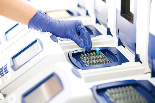
Within the next decade or two, qPCR will be the most sensitive, specific, and also the most reliable method for molecular analysis in clinical settings. [vkovalcik/iStock]
GEN Exclusive: Reminiscing with Kary Mullis
GEN: How did the idea of PCR come to you?
Mullis: At first I was not even looking for a method to amplify DNA. I was working on techniques for using oligonucleotides to investigate single base pair mutations that might be present at a particular location on DNA that a doctor might have isolated from an unborn child to see if the child had a genetic disease.
Back then, to do something like this, you had to clone the DNA. Sometimes this could take several weeks. This would obviously pose a problem to a woman who needed to decide as soon as possible if she was going to have an abortion.
But one night, while driving on a road in California, I was thinking about new ways to use oligos to sequence DNA more effectively to study genetic diseases. I decided that two oligos would be better than one because each oligo could be directed to each strand of the DNA. This thought process put me on the path to discovering PCR.
GEN: There are a number of life science areas you could have chosen to study. What was it about oligos and DNA synthesis that specifically caught your attention
Mullis: The same thing that influenced Ron Cook, who founded Biosearch, which manufactures oligonucleotides. He and I were both peptide chemists at the University of California in San Francisco. One day we went to a seminar and learned that bacteria could now make peptides. I remember I looked at Ron and he looked at me, and the unspoken message was, “We are about to be replaced by bacteria.” So we decided we should start making oligonucleotides instead.
Ron eventually started working on a machine that would manufacture oligonucleotides, which later to drove me to invent PCR.
GEN: What were some of the first applications for PCR?
Mullis: Right down the hall from my lab at Cetus, there was a group headed by Henry Ehrlich. Randy Saiki worked in that lab. Randy and his colleagues were trying to come up with an in vitro diagnostic for sickle cell anemia that would work on small DNA samples that they could get from a fetus. I was able to amplify the sickle cell gene with PCR. Tom Caskey’s lab at Baylor later used PCR in their work on Duchenne muscular dystrophy.
GEN: What did it feel like to win a Nobel Prize?
Mullis: It changed my life. When a country and its king gives you such an award that kind of impresses the heck out of you. All of a sudden you are a celebrity and not just a grant scientist. Everybody in the world wants you to come to their university and give a lecture. You feel like a rock star, for at least a few months.

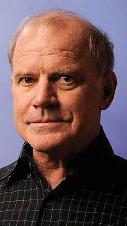
Kary Mullis, Ph.D., received a Nobel Prize in chemistry in 1993 for his invention of the polymerase chain reaction (PCR). This technology, which was discovered thirty years ago and which GEN is celebrating in this special section, has transformed and revolutionized life science research. Dr. Mullis recently granted GEN an exclusive interview.
Mikael Kubista, Ph.D. ([email protected]), is CEO and founder of TATAA Biocenter in Goteborg, Sweden.


