February 1, 2013 (Vol. 33, No. 3)
High-content screening (HCS) has emerged as a unique approach that combines the power of automated image analysis with fluorescence microscopy in detecting various cellular processes and interactions.
This approach has been supported by almost a decade of efforts in improving methods for the quantification of signals and management of image data files.
At CHI’s recent “High-Content Analysis” conference, several scientists and computer software developers presented novel approaches to high-content screening in different scenarios and platforms.
High-content screening experiments by their nature generate a tremendous amount of information in images and numerical results, according to Oliver Leven, Ph.D., head of professional services of Genedata Screener®.
“Each well in a microtiter plate contains hundreds or thousands of cells, and the goal of high-content screening is to capture these cells and to quantify their response to the changed experimental conditions as expressed by their phenotypes. Typical experiments range from four 96-well plates to hundreds of 384-well plates. Combining the results from multiple wells enables researchers to study the potency of new chemical substances or to quantify the damage to cells after drug administration.”
To support such experiments, Genedata has thus developed a data analysis workflow system that consolidates image-data gathered from multiple instruments, allowing researchers to import data into a single platform.
“To look at one screen for the analysis of dose-response curves and then go to another computer—or just change software to compare the underlying images is a time-consuming and painful activity for a scientist,” explained Dr. Leven. “The Genedata Screener image workflow system has been designed as a scalable platform that can handle up to 400 cellular features generated from different instruments and analyze them side by side in a single computer screen with easy access to images.”
An in-depth analysis of HCS experiments with thousands of images is often prevented due to the difficulties in accessing the enormity of available information.
“One common stumbling block for phenotypic screening assays involves knowing where your cells are after running a microplate-based experiment. Every researcher wants to interpret files from yesterday’s experiment, see the raw data, and compare this to today’s experiment. However, once you scale-up your experiments to process 50 96-well plates, data workflow becomes a problem,” Dr. Leven further explained.
“Often the scientist spends more time on obtaining the information than on analysis itself.” He said that Genedata Screener supports reliable throughput information and full access to all information enabling the processing of the data that gives value to each well analyzed.
High-content screening has also been employed in toxicity assessment of new drugs. According to Evan Cromwell, Ph.D., director of research at Molecular Devices, phenotypic screening assays are useful in identifying drugs that could damage certain types of cells.
“Toxicology is generally difficult to predict, even in animal models and humans, and thus high-content screening assays using stem cell-derived cardiomyocytes, hepatocytes, and neurons can facilitate the determination of complex responses and phenotypes, for example, viability and mitochondrial damage, due to exposure to specific drugs,” Dr. Cromwell said.
In addition, high-content screening also allows scientists to multiplex information from other assays for specific cellular activities, including caspase activation, DNA damage, and changes in nuclear shape. Cell morphology, adhesion, and spreading are indicators of toxicity and they need to be accurately characterized by software analysis.
Together with the efforts of other scientists at Molecular Devices, the success of phenotypic screens is based on the development of instrumentation that allows them to capture images that could be subjected to statistical image data analysis.
“These algorithms for analysis of cell-based assays allow us to identify which drugs are safe or toxic, and potentially predict mechanisms of action,” explained Oksana Sirenko, Ph.D., research scientist at the firm. “We have done proof of concept for cardiotoxicity and hepatotoxicity that shows such assays can be predictive, yet also face certain challenges.”

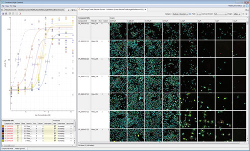
Genedata Screener for HCS supports dose-response curve (DRC) fitting: efficient fitting and display of DRCs (upper left), and overview of phenotypic changes across the dilution series (right). Support is also provided to fit multiple features per compound.
Cell Migration
The technology of high-content screening has also been applied to the concept of cell migration in embryonic development and cancer. Michael Prummer, Ph.D., scientist for high-content screening at F. Hoffmann-La Roche, said that phenotypic screens are important in identifying cellular changes that are well beyond classical biochemical assays.
“We look at the full complexity that one cell can offer, and by using high-content screening we are now capable of generating a wealth of information at high depth and high speed. We examine cell migration to determine the potency of drugs in vitro, with the aim to predict how these compounds affect the cell migration machinery in vivo,” he explained.
Dr. Prummer also discussed that by targeting the activity of cell migration, it is possible to screen hundreds of unknown proteins using high-content screening. “Unlike target-based screening that starts with a well-characterized protein target and finding compounds that modulate its functions, phenotypic screening focuses on a cellular activity, morphology, or state—which in our case is cell migration—and we test various compounds to find a lead structure that can result in a change in cell mobility.”
He further explained that this approach circumvents some of the technical hurdles that are associated with traditional assays on cell migration, such as the scratch assay. “We can now observe cells moving in a plate on an individual basis, perform image analysis, and quantify the extent of motion of each cell.”
Reliability and Flexibility
High-content screening has also been further improved in terms of image quality as it relates to image reliability, or how closely that image represents the biology of a sample. “For high-content analysis, the quality of your data is only as good as the reliability of the detector, and it is not simply based on resolution and sensitivity,” said Audra Ziegenfuss, technical product manager of high-content products at Thermo Scientific.
“Due to the new X1 CCD camera chip’s architecture and read-out, it is highly linear (
Dr. Ziegenfuss also emphasized that researchers need flexibility during high-content screening. “As you conduct a high-content screening assay, you want to know that your camera can be used for various imaging modes, and together with the features of the sensor and the software, one can orchestrate together a high-quality image that gives you statistically relevant data. With advances in microarray technology and binning, quantum efficiency can be maximized up to 75% without sacrificing resolution,” she added.
“For confocal applications, which can often result in only 3–16% of the total fluorescence emission during imaging, and live cell applications sensitive to issues of phototoxicity to cells, this is important.”
The development of algorithms for application to high-content screening has also further enhanced the capacity for analyzing cellular activities. “Most of researchers are throwing away so much data due to limitations in finding and characterizing cells,” explained David Novo, president and CEO of De Novo Software.
“By using population-based analysis of cells, you can characterize each type of cell in terms of size, nuclear staining, and shape, and consequently create gates based on these parameters. This visual verification of cell parameters allows the scientist to identify differential effects of a drug in each well of a microtiter plate.” He also explained that the use of algorithms in image data analysis allows the researcher to plot numerical values based on visual characteristics of cells.
“What used to be single-color experiments in a single well can now be performed using multiple colors, yet increasing the capacity for image analysis at a single-cell level. The De Novo Cell Profiler offers the power of screening thousands of cells per well using several fields of view. This software technology generates gates in which one can perform statistical analysis, preventing the researcher from dumping most of the data gathered from an experiment.”
Into the Clinic
Profiling phenotypic effects of drugs for potential clinical use has also relied on the information generated by high-content analysis platforms. “Our ongoing collaborative project with Genentech involves looking at the response of Cytiva cardiomyocytes to kinase inhibitors to gain information on drug toxicity and mechanism of action,” explained Nick Thomas, Ph.D., principal scientist at cell technologies, GE Healthcare.
“There are drugs that may have off-target effects on other cells, such as the heart, and thus, our high-content screening assay allows us to track functional changes in mitochondria. We also look into calcium release and homeostatis as a cytotoxic response of cells to stress, including that imparted by drugs.”
Dr. Thomas further discussed that changes in the morphology and membrane potential of mitochondria and calcium homeostasis provide information on acute and chronic drug exposures, helping them identify potentially toxic drugs. “It is also possible that a certain drug be administered using a different dosing and this information can be derived from high-content screening assays, where cells are exposed to a range of concentrations at different time points.”
According to Dr. Thomas, high-content screening is an automated differential screen that provides the researcher the breadth and depth of information needed in drug toxicity studies.
“This valuable information we get from high-content analysis also helps in prioritizing compounds for drug development and potential release to the market. Even at the early stages of drug development, once you determine by high-content screening that a certain compound is affecting mitochondria and induces cellular toxicity, then you can engineer changes to the compound to reduce toxicity or look at modifying its concentration or timing of drug administration,” he explained. His group also envisions building a database of new drugs with signatures of known clinical toxicities.

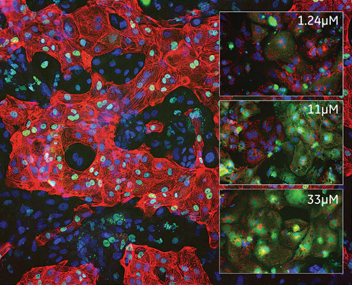
GE Healthcare CytivaT human stem cell derived cardiomyocytes: Inset images from HCA cardiotoxicity assay show cells exposed to increasing concentrations of an anticancer drug showing disruption to mitochondrial integrity (red) and calcium homeostasis (green).


