Kevin Mayer Senor Editor Genetic Engineering & Biotechnology News
One way or another, scaffolds, stem cells, and biological factors come together to recapitulate organ development in the lab.
Although a body, no less than a car, may eventually need replacement parts, surgeons cannot simply place an order at the Organ Zone, and they never will, for biological organs, unlike car parts at the Auto Zone, cannot be mass produced. The old assembly line—technology’s best answer to closing gaps in supply and demand—fails patients on organ transplant waiting lists because biological organs are simply too complex. Each organ is unique. What’s more, it has a short shelf life, so stocking inventory is out of the question.
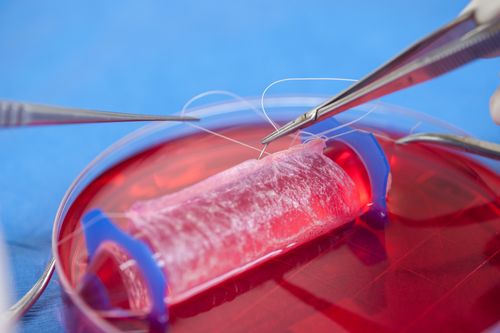
One alternative to waiting for a donor organ is to handcraft one. Doing so is as inconvenient at it sounds. First, you need a scaffold. This, too, can come from a donor, a cadaver even, provided all the cells are removed, leaving only extracellular matrix consisting of collagen or cartilage. Or porous plastic can serve the purpose, or decellularized animal tissue. In any case, once you have a scaffold, you can painstakingly seed it with stem cells while making sure it is continuously incubated.
Naturally, the stem cells would come from the patient in need of the replacement organ. By eliminating donor cells and using a patient’s own cells, you can avoid immune rejection of the organ once it has been implanted.
Some degree of immune response, however, is necessary. Such a response, at the graft/host interface, helps stimulate signaling mechanisms, changes in gene expression, and cell-mediated repair mechanisms. By means not yet understood, the body’s recognition of the replacement organ, or even the molecular debris it may shed, makes it possible for the new organ to become integrated with the patient’s tissues. Even blood vessels and capillaries form to nourish the scaffold-embedded cells and carry away wastes, provided a barrier of scar tissue isn’t allowed to form.
To date, this approach has been used to accomplish relatively simple repairs in human patients. For example, scaffolds fashioned from cadaver materials and plastics have been seeded with stem cells to create new windpipes. Thin sheets of animal tissue, from pigs, were used to support the growth of new muscle in injured veterans. Collagen membranes have been used to engineer human cartilage grafts from patients’ own nasal septum cartilage cells to successfully reconstruct nostrils. And specially constructed biodegradable molds have been seeded with cells from patient biopsies to create artificial bladders and vaginas.
Replacement Parts Show Signs of Long-Term Success
Follow-up studies have been encouraging. For example, in April 2014, one year after surgery, all five recipients of reconstructed nostrils said that they were satisfied with their ability to breathe, as well as the cosmetic appearance of their nose, and did not report any local or systemic adverse events. One of the scientists involved, Ivan Martin, Ph.D., from the University of Basel in Switzerland, said that this success “opens the way to using engineered cartilage for more challenging reconstructions in facial surgery such as the complete nose, eyelid, or ear. The same engineered grafts are currently being tested in a parallel study for articular cartilage repair in the knee.”
The researchers responsible for the laboratory-grown bladders and vaginas have conducted even lengthier follow-ups. Anthony Atala, M.D., director of the Institute for Regenerative Medicine at Wake Forest University School of Medicine, and colleagues noted that bladders implanted in patients aged 4 to 19 years old showed improved function over time—with some patients being followed for more than seven years.
Wake Forest also reported on the long-term success of tissue-engineered vaginal organs implanted in four women, aged 13 to 18 years, with a condition known as Mayer-Rokitansky-Küster-Hauser syndrome that causes the vagina to be underdeveloped or absent. In April 2014, eight years after transplantation, the organs continue to function as if they were native tissue and all recipients are sexually active, report no pain, and are satisfied with their desire/arousal, lubrication, and orgasm.
According to Dr. Atala, “Yearly tissue biopsy samples show that the reconstructed tissue is histologically and functionally similar to normal vaginal tissue. This technique is a viable option for vaginal reconstruction and has several advantages over current reconstructive methods because only a small biopsy of tissue is required, and using vaginal cells may reduce complications that arise from using nonvaginal tissue (segments of large intestine or skin) such as infection and graft shrinkage.”
In a comment published in the Lancet and linked to both the reconstructed nostril and lab-grown vagina studies, Martin Birchall, M.D., of UCL Ear Institute, London, U.K., wrote, “These authors have not only successfully treated several patients with a difficult clinical problem, but addressed some of the most important questions facing translation of tissue engineering technologies.
“The steps between first-in-human experiences such as those reported here and their use in routine clinical care remain many, including larger trials with long-term follow-up, the development of clinical grade processing, scale-out, and commercialization,” Dr. Birchall continued. “However, these hurdles are common to all potentially disruptive technologies, and many countries now have large translational income streams, engaged biotech companies and streamlined regulatory processes that may reduce the time to routine use significantly.”
Mass-Customizing Organs
While optimism does seem to be in order, this approach to tissue engineering, with its arduous and time-consuming shaping of scaffolds and pipetting of stem cells, has something of a preindustrial, handicraft feel to it. Moreover, it seems inherently resistant to the sort of top-down optimization you might see in an industrial setting. Mass production would seem to be out of the question. But what about mass customization?
Mass customization refers to a kind of bottom-up process that relies on computer-aided design (CAD) tools and rapid prototyping platforms such as 3D printers. Although 3D printers have been around for the last two decades, fabricating all sorts of objects out of protoplastics and other “inks,” they have only recently been shown capable of extruding or spraying biological material such as living cells. Since it was learned that more than 90% of bioprinted cells manage to survive the rigors of bottom-up processing including the curing of liquid or gelatinous gels, which serve as cell carriers until they harden into scaffolds, researchers interested in generating 3D replacement organs have been pressing the “print” button. If they succeed, they will create an industry that has the distinction of skipping the mass production phase of industrial development.
That may seem too large a chasm to cross. The other side may be reached, however, partly thanks to insights gleaned from top-down systems. For example, it is already well understood that printing an organ won’t be as simple as dropping cells and supporting materials in the right place and calling it a finished product. An organ grows and develops over time amidst a storm of signals and signal responses, and these depend on environmental cues and cellular sensitivities and propensities for self-organization.
Exactly how all these interactions can be orchestrated is unclear, but researchers, undaunted, are pressing forward. Some are using 3D printers to create grow artificial scaffolds, which are then seeded with stem cells much like scaffolds of the handcrafted variety. Other researchers are laying down cells and artificial matrix materials simultaneously. Finally, some researchers are intent on using only biological materials, avoiding artificial materials entirely. (The logical extreme of this approach might be entirely chemical, stimulating the patient’s tissues to regenerate on their own, skipping the generation and implantation of a bioficial organ altogether.)
Another complication is that 3D printing technology still lacks the resolution to lay down complex, interconnected capillary networks, limiting the depth of bioficial tissues. Nonetheless, researchers are working out various expedients, including the laying down of sacrificial threads. Once such threads biodegrade or melt or dissolve away, the spaces they leave behind can serve as conduits for blood vessels.
New Developments
While the ultimate goal is to create implantable and immunologically compatible organs, bioprinting technology’s near-term goals are somewhat less ambitious. Recent announcements highlight progress on biosensors, “organ-on-a-chip” devices (such as Organovo’s liver chip, which is being developed for drug toxicity testing and other applications), 3D tissue models of disease, patches to repair largely intact body structures, and (apologies if you’ve heard this one already) the $300,000 hamburger.
In just the past few months, multiple research groups have reported diverse bioprinting advances:
• Heart Disease on a Chip: Harvard scientists have merged stem cell and organ-on-a-chip technologies to grow, for the first time, functioning human heart tissue carrying an inherited cardiovascular disease. The scientists published their work May 11 in Nature Medicine, in an article entitled “Modeling the mitochondrial cardiomyopathy of Barth syndrome with induced pluripotent stem cell and heart-on-chip technologies.”
Instead of producing single heart cells in a dish, the scientists grew stem cells on chips lined with human extracellular matrix proteins that mimic their natural environment, tricking the cells into joining together as they would if they were forming a diseased human heart. The engineered diseased tissue contracted very weakly, as would the heart muscle seen in Barth syndrome patients.
In the article, the authors concluded that their study “provides new insights into the pathogenesis of Barth syndrome, suggests new treatment strategies, and advances iPSC-based in vitro modeling of cardiomyopathy.”
• Microrobotic Assembly Technique: Researchers at Brigham and Women’s Hospital and Carnegie Mellon University introduced an untethered magnetic microrobotic technique for the precise construction of individual cell-encapsulating hydrogels (such as cell blocks). In an article that appeared February 10 in Nature Communications, the authors explained that their microrobot, which is remotely controlled by magnetic fields, can move one hydrogel at a time to build complex structures.
This capability is critical in tissue engineering, which seeks to emulate how human tissue is composed of different types of cells at various levels and locations. In natural and laboratory-grown tissues alike, the location of the cells impacts how larger structures will ultimately function. If numerous microrobots were to be used together in bioprinting, tissue engineers might flesh out complex designs with hydrogel-encapsulated cells better able to maintain their vitality and ability to proliferate.
• Ultrathin Collagen Matrix Biomaterial for Organ Chips: A team of researchers from the Center for Engineering in Medicine at the Massachusetts General Hospital have demonstrated a new nanoscale matrix biomaterial assembly that can maintain liver cell morphology and function in microfluidic devices. The researchers, led by Martin Yarmush, M.D., Ph.D., published their findings March 24 in Technology.
“This is a clever combination of the well-known layer-by-layer deposition technique for creating thin matrix assemblies and collagen functionalization chemistries that will really enable complex liver microtissue engineering by replicating the physiological cues that maintain the state of liver cell differentiation,” said Dr. Yarmush. “The ultrathin collagen matrix biomaterial and its ability to keep liver cells functional for longer periods of time in chip devices will undoubtedly be a useful tool for creating liver microtissues that mimic the true physiology of the liver, including cell and matrix spatial geometries.”
• Growing Human Cartilage in the Lab: The general approach to cartilage tissue engineering has been to place cells into a hydrogel and culture them in the presence of nutrients and growth factors and sometimes also mechanical loading. But using this technique with adult human stem cells has invariably produced mechanically weak cartilage.
In hopes of producing stronger cartilage, researchers at Columbia Engineering came up with a new approach: inducing mesenchymal stem cells to undergo a condensation stage as they do in the body before starting to make cartilage. The researchers, led by Gordana Vunjak-Novakovic, Ph.D., reported that they were able to grow fully functional human cartilage. In particular, as detailed in a paper that appeared April 28 in the Proceedings of the National Academy of Sciences, the tissue-engineered cartilage approached native cartilage in terms of its lubricative properties and compressive strength.
In another laboratory-grown cartilage advance, researchers at the University of Pittsburgh School of Medicine reported that they had used a bioprinting technique to create the first example of living human cartilage grown on a laboratory chip. The feat was described April 27 at the Experimental Biology 2014 meeting. The presentation, delivered by Rocky Tuan, Ph.D., director of the Center for Cellular and Molecular Engineering, was called “Biomimetic scaffolds and natural matrices for stem cell-based tissue engineering and modeling.”
Housing 96 blocks of living human tissue four millimeters across by eight millimeters deep, the chip faithfully represented the bone-cartilage interface. It could serve as a test-bed for researchers to learn about how osteoarthritis develops and develop new drugs. “With more testing, I think we’ll be able to use our platform to simulate osteoarthritis, which would be extremely useful since scientists really know very little about how the disease develops,” predicted Dr. Tuan.
Dr. Tuan also reported progress toward his ultimate vision: giving doctors a tool they can thread through a catheter to print new cartilage right where it’s needed in the patient’s body. According to
Dr. Tuan, this vision may be realized via a technique that extrudes thin layers of stem cells embedded in a solution that retains its shape and provides growth factors. This solution is an injectable, biodegradable methacrylated gelatin-based hydrogel that is capable of rapid gelation via visible light-activated crosslinking in air or aqueous solution. The mild photocrosslinking conditions, which avoid the use of UV light, permit the incorporation of cells during the gelation process.
• Growing Vascularized Tissues in the Lab: A new bioprinting method developed at the Harvard School of Engineering and Applied Sciences (SEAS) and the Wyss Institute for Biologically Inspired Engineering at Harvard University creates intricately patterned, three-dimensional tissue constructs with multiple types of cells and tiny blood vessels. To print 3D tissue constructs with a predefined pattern, the researchers needed functional inks with useful biological properties, so they developed several “bio-inks”—tissue-friendly inks containing key ingredients of living tissues.
One ink contained extracellular matrix, the biological material that knits cells into tissues. Another ink contained both extracellular matrix and living cells. Yet another ink was used to create blood vessels. It was developed to melts as it cools, rather than as it warms. This allowed the scientists to print an interconnected network of filaments that they could then melt by chilling the material, which was ultimately suctioned out to create a network of hollow tubes, or vessels.
The scientists, led by Jennifer Lewis, Ph.D., printed 3D tissue constructs with a variety of architectures, culminating in an intricately patterned construct containing blood vessels and three different types of cells—a structure approaching the complexity of solid tissues. Moreover, when they injected human endothelial cells into the vascular network, those cells regrew the blood-vessel lining.
• The Motherboard of All Organ Chips: Organ chips, microfluidic devices that amount to compact, three-dimensional cell culture versions of real organs, already exist for skin, cartilage, bone, gut, artery, heart, and kidney. But now researchers, with support from various defense agencies, are intent on linking them together to create a body on a chip. Such a device would be analogous to a computer motherboard; however, instead of electrical subassemblies, this device would accept organ chips as plugins. The connections would not be electrical but microfluidic. A bloodstream in miniature would have to be contrived.
One body chip project is being led by Rashi Iyer, Ph.D., senior scientists at Los Alamos national Laboratory (LANL). The project, called ATHENA (for the Advanced Tissue-engineered Human Ectypal Network Analyzer), is developing four human organ constructs—liver, heart, lung, and kidney. Each organ component will be about the size of a smartphone screen, and the whole ATHENA body of interconnected organs, jocularly referred to as “homo minutus,” would fit neatly on a desk.
Project components are being divided among various institutions. For example, a blood mimic to sustain the four organ constructs is being developed by LANL and Vanderbilt University in collaboration with CFD Research Corporation.
Another possibility for microfluidic blood is being explored by researchers at the Wyss Institute. These researchers have developed a bone marrow-on-a-chip that they say reproduces the structure, functions, and cellular makeup of bone marrow. These findings appeared May 4 in Nature Methods, in an article entitled “Bone marrow-on-a-chip replicates hematopoietic niche physiology in vitro.”
While the researchers emphasized their work’s immediate applications, which include drug testing, they also note that their chip could generate blood cells that could circulate in an artificial circulatory system to supply a network of other organs-on-chips. According to a release that cited the Nature Methods article, the Defense Advanced Research Projects Agency (DARPA) is providing funds to the Wyss Institute to develop an interconnected network of 10 organs-on-chips to study complex human physiology outside the body.


