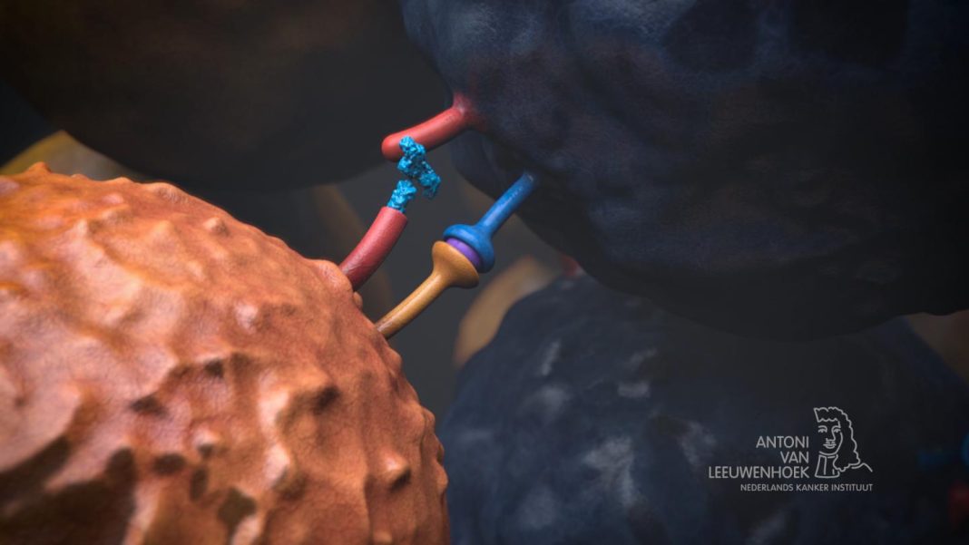
Myocarditis—inflammation of the heart muscle—is a rare, but potentially fatal complication of immune checkpoint inhibitor (ICI) cancer immunotherapy. Researchers at Vanderbilt-Ingram Cancer Center have now discovered that the mechanism for this serious adverse event involves T-cells recognizing the cardiac antigen α-myosin. The team suggests that findings from their experiments in mice, and from studies using blood cells taken from patients with ICI-related myocarditis (ICI-MC) could help scientists identify biomarkers so that at risk patients can be recognized and strategies developed to allow them to tolerate the immunotherapy.
Headed by Justin Balko, PharmD, PhD, Ingram Associate Professor of Cancer Research at Vanderbilt-Ingram Cancer Center, the researchers described the research in Nature, in a paper titled “T cells specific for α-myosin drive immunotherapy-related myocarditis.” In their report the team concluded that identification of α-myosin as an autoantigen may “ … guide the identification of biomarkers to predict which patients are at higher risk for myocarditis, such as surveillance of peripheral α-myosin-reactive T cells or identification of pre-existing autoantibodies.”

Clinicians don’t yet have a clear understanding of why immunotherapy-related myocarditis occurs in some patients. Although early treatment with steroids can improve survival chances, a more effective treatment is urgently needed. “… preventing, diagnosing and treating irAEs are urgent clinical challenges,” the scientists wrote. “Currently, clinically actionable biomarkers of response and toxicity are limited, and the mechanistic basis of irAEs is poorly defined.”
In 2016, the Balko group first described two melanoma patients treated with immunotherapy who developed myocarditis, and the researchers performed some early studies linking the activity of T cells in the heart to the condition. “Subsequently, working with the Nobel laureate James P. Allison, PhD from MD Anderson Cancer Center in Houston, we helped characterize a mouse model that seemed to replicate what we had observed in patients,” Balko noted. “Using that same model, together with oncologist Douglas Johnson, MD, MSCI at Vanderbilt and co-corresponding author Javid Moslehi, MD at UCSF, we were able to pinpoint the mechanism of why it occurs—and importantly—translate this back to patients. This discovery represents the next important step to making these often-effective therapies safer in patients.”
For the newly reported studies the research team obtained cardiac samples and peripheral blood from three patients who had suffered severe myocarditis after being treated with immune checkpoint inhibitors. These samples were analyzed after the team replicated immunotherapy-related myocarditis in a mouse model. “Pdcd1–/–Ctla4+/– mice recapitulate clinicopathological features of ICI-MC, including myocardial T cell infiltration,” they explained.
The researchers sequenced individual T cells invading the heart during myocarditis in the mouse models to reconstruct their receptors. These T cell receptors (TCRs) were then screened against peptides to determine specificity. “We showed in our mouse model that myocarditis is characterized by cytotoxic CD8+ T cells with highly clonal TCRs, and that CD8+ cells are necessary for the development of myocarditis,” the authors explained. “Three of the most clonal TCRs, derived from independent mice, recognized α-myosin epitopes.” When the researchers then analyzed the human samples, they found that the three patients also had reactive T cells to α-myosin, which is expressed only in heart and skeletal muscles. “We establish CD8+ T cells as necessary for disease and identify α-myosin as a cognate antigen for the most abundant TCRs in myocarditis,” the team stated. “Furthermore, we extend these findings to human disease and show that α-myosin-expanded TCRs are present in inflamed cardiac and skeletal muscle in patients with ICI-MC.”
“The extension of our findings from the mouse model to human patients was a key part of our work,” commented first author Margaret Axelrod, PhD, a Vanderbilt Medical Scientist Training Program student who completed her PhD in the Balko Lab.” These results show how useful it is to have a mouse model where you can make an initial discovery and use that to understand something about human disease. Our data show that α-myosin is a disease-relevant autoantigen in patients with immunotherapy-related myocarditis. We hope this mechanistic understanding of this often-deadly complication can pave the way toward making immunotherapy safer for patients.”
The study is the first to identify the role of α-myosin in the mechanism of heart complications from immune checkpoint inhibitors, and is also among the first to identify a candidate autoantigen for an immunotherapy toxicity in humans, the scientists claim. “These studies underscore the crucial role for cytotoxic CD8+ T cells, identify a candidate autoantigen in ICI-MC and yield new insights into the pathogenesis of ICI toxicity … Knowledge of the most relevant disease antigens may facilitate antigen-directed approaches to suppress inflammation without compromising on antitumour efficacy such as tolerogenic vaccines.”
Balko added. “While autoreactive T cells are the presumed mechanism for many toxicities to immunotherapies, tracing the condition back to a specific T cell receptor (often unique to each patient) and antigen source (drawing from tens- to hundreds- of thousands of potential antigens in the human body) is a daunting task.”


