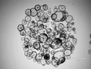
 Three-dimensional (3D) cell culture overcomes many shortcomings of two-dimensional (2D) culture, mainly through the introduction of biological relevance. Cells in three dimensions “see” and “feel” neighboring cells, interact with them through chemical messaging, and reprise physiologic events much more faithfully than cells restricted by the physical and mechanical constraints of standard cultureware.
Like any emerging technology, 3D cell culture has advanced in several directions, most notably as 3D suspended cultures, known as spheroids and organoids. As 3D culture has grown in popularity, the terms “spheroid” and “organoid” have been used interchangeably; however, the differences between these culture types are significant.
So, what are the differences?
Spheroids and organoids both contain multiple cells, typically suspended within a droplet or microwell. Spheroids are simple clusters of broad-ranging cells collected and cultured from various tissues, for example, from an organ or, by way of a biopsy, from a diseased tissue or tumor. Spheroids contain a representative sampling of the tissue’s cell types, usually without selection or curation for specific cells. Sometimes the sample contains one cell type, sometimes more than one. Spheroids are known for being easier to work with, as they do not require physical scaffolding to form. This, of course, can have its downsides. Spheroids are simpler models that do not self-organize or have polarity and are thus not always representative of the more complex in vivo environment.
As their name implies, organoids are complex clusters of organ-specific cells designed to mimic the original tissue such as the skin, stomach, liver, or bladder. Unlike spheroids, which form from tissue samples containing any number and type of mature cells, organoids arise from tissue-specific stem cells, or progenitor cells, harvested from various organs, such as the brain or liver. Organoids can also be produced from induced pluripotent stem cells. Once provided with an appropriate extracellular matrix and treated with biochemical stimuli (specific for the eventual target cells and the tissue of interest), these progenitor cells expand, differentiate, and self-assemble into microscopic cultures that recapitulate one or more critical aspects of the original tissue.

Collagen and Corning® Matrigel® matrix are two typical scaffold materials you can find in a laboratory. Matrigel, a gelatinous protein mixture derived from Engelbreth–Holm–Swarm (EHS) mouse sarcoma cells, contains several proteins, carbohydrates, and growth factors that are also present in tumor microenvironments. With more than 10,000 scientific citations,1 Matrigel is the most-used extracellular matrix for organoid formation. The matrix is so common that its trademarked name has almost become genericized. (Genericized names include Kleenex, the popular facial tissue.)
A search engine query for “commercially available organoids” brings up dozens of examples. Many more are easily accessible through established protocols, which include recipes for organoid models of liver, heart, pancreas, brain, gastrointestinal tract, kidney, and (more recently) lung. Lung or human airway organoids are suitable for the study of infectious human respiratory diseases.
Spheroid applications
Although spheroids lack organoids’ engineered complexity, their 3D structure puts them miles ahead of conventional 2D cell culture models, particularly for drug screening. Interestingly, spheroids were initially developed in the 1970s to study the effects of radiotherapy on human tumors—a prophetic nod to their current use.
Tumor-derived spheroids reproduce both the specific tumor cell types—with all their abnormalities—and the physical–mechanical environment that makes tumors so difficult to treat—a factor totally absent when assaying cancer cells plated in 2D. Notably, spheroids permit the direct study of the tumor microenvironment, which is a major factor in how cancers respond to drug treatment.
Scientists have used tumor spheroids to study the cytotoxic effects of chimeric antigen receptor (CAR) T cells—such as the KILR® Cytotoxicity assay developed by DiscoverX. When CAR T cells are grown in KILR-transduced tumor spheroids, scientists have the ability to form, culture, and assay in the same spheroid microplate. This theme of high-throughput 3D assays in which multiple readouts are obtainable, recurs over and over in any discussion of 3D cell culture.
Organoid applications
As mentioned, organoids represent the in vivo environment more faithfully than spheroids, but producing organoids involves a more complex process, and maintaining them is more difficult as well. Academic groups use organoids in the study of basic biology,2 organ development, and tissue morphogenesis. Further along the application curve, organoids serve as disease models, which allow direct comparisons between healthy and diseased tissues. Organoids provide a platform for assessing, in one experiment, differences among various cells in responses to toxins, drugs, or environmental stresses. Far in the distant future, one could envision organoids, or similarly configured engineered in vitro tissues, as replacements for failing kidneys or livers.
Organoid methodology in its present form would not be possible without the pioneering work of Hans Clevers, MD, PhD, professor of molecular genetics, University Medical Center Utrecht and Utrecht University. In 2009, he described methods for growing and expanding human adult stem cell–derived epithelial organoids. Under Clevers’ direction, Hubrecht Organoid Technology, a Netherlands-based company, has produced and validated organoid models for cancers of the colon, breast, lung, liver, ovary, bladder, and pancreas, and for other diseases. Clevers’ discovery allowed for the expansion of adult stem cell–derived organoids in a genetically stable form. Most importantly, it allowed organoids to expand indefinitely, like tumor cells, but without the genetic abnormalities inherent in cancer cells and, in fact, without the need for any genetic modification.
Conclusion
2D cell cultures lack the structure, function, dimensionality, cellular diversity, and cell–cell interactions that make living tissue unique, particularly for the study of higher-order biology.
Much has been written about drug development failures, their causes, and the heavy financial toll they impose on pharmaceutical companies. A significant number of these failures may be attributed to preclinical strategies relying on 2D cell culture–based assays which, after all, are expected to predict safety and efficacy in humans—not in Petri dishes. Furthermore, experts recognize the limitations of 2D cell culture as a contributing factor to these failures.3
3D cultures involve significantly more attention and resources to get right, as opposed to the simple 2D plating of cells. Adopting spheroids or organoids in a drug screening program also requires a new way of thinking about cell-based assays. In return for this effort, 3D cell cultures provide an opportunity for in vitro assays that may better mimic what is happening in vivo.
Download Infographic or Request the Poster
Elizabeth Abraham, PhD, is senior product manager at Corning Incorporated, and Hilary Sherman is senior applications scientist and Audrey Bergeron is applications scientist II at Corning Life Sciences.
References
1. Slater K, Flaherty P. Everything you ever wanted to ask about Corning® Matrigel® Matrix. Cell Culture Dish. Published October 2, 2017. Accessed September 29, 2020.
2. Lo YH, Karlsson K, Kuo CJ. Applications of organoids for cancer biology and precision medicine. Nature Cancer 2020; 1: 761–773.
3. DePalma, A. INSIGHTS on 3D Cell Culture and Organs-on-Chips. Lab Manager. V2BAGpEwhhH. Published April 6, 2016. Accessed September 29, 2020.

