September 15, 2014 (Vol. 34, No. 16)
Testing Drug Efficacy by Rapid Size Profiling Over Time with Tumor Microtissues
It is commonly accepted that tumor sensitivity or resistance to chemotherapeutic agents is not only genetically determined, but also driven by the tumor microenvironment. Metabolic gradients and the extracellular matrix can influence nutrient availability to various segments of a solid tumor, and also limit the ability to administer an effective dose of chemotherapeutic agent. These biological and therapeutic gradients result in the formation of phenotypically different tumor cell subpopulations exposed to a variety of therapeutic to subtherapeutic drug doses in vivo.
Tumor size is the most frequently used in vivo endpoint when assessing antitumor efficacy in animal xenograft models, whereas proliferation is the more typically evaluated growth endpoint in vitro using two-dimensional (2D) monolayer cultures. Such 2D in vitro assays frequently fail to correlate with in vivo observations, owing to the inability of 2D cultures to recapitulate the native tumor microenvironment described above. Three-dimensional (3D) tumor microtissues, or multicellular tumor spheroids, are considered a more representative, organotypic model for assessment of tumor growth. They contain layers of cells that exhibit more in vivo-like size- and gradient-dependent proliferation and viability profiles.
For example, more proliferative cells tend to cover the outer oxygen and nutrient-rich layers of the spheroid, whereas the nutrient-restricted inner cells tend to display a more quiescent or even necrotic phenotype, as the spheroid diameter increases. Additionally, the ability to monitor spheroid growth in terms of size provides a similar unit of measure with which to make a direct in vitro to in vivo comparison.
Although the benefits of 3D culture models have been widely acknowledged, their widespread implementation in drug discovery screening efforts has been slowed by the lack of appropriate companion assays and instrumentation to maximize their value. Imaging spheroids to monitor their growth using conventional or even automated microscopes is a slow, low-throughput process. High-content imaging systems overcome some of these throughput issues, but are comparatively expensive, and still suffer from slow image capture/processing, and image analysis software that has not been optimized for 3D spheroids.
Although adaptable to higher-throughput plate readers, biochemical assays that monitor viability can effectively correlate to spheroid size, but are often lytic or otherwise destructive in nature, requiring multiple replicates to be run for longitudinal studies.
For this study, we aimed to improve the throughput of monitoring tumor spheroid growth in response to anticancer drugs in vitro using the Dainippon SCREEN Cell3iMager, a rapid, robust brightfield imager for measuring spheroid size and morphology.
Rapid Assessment of Spheroid Size and Morphology
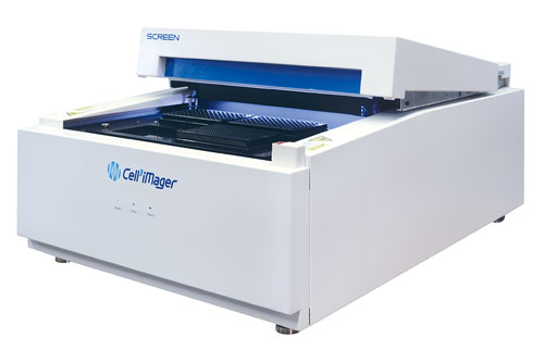
The Cell3iMager (Figure 1) facilitates analysis of spheroids by fast, parallel scanning in a brightfield, allowing determination of spheroid count per well, diameter, area, pseudo volume, and loss of circularity in individual spheroids. Image capture is rapid, requiring less than 1 minute per plate at 2,400 dpi (10.6 μm/pixel). The 4-plate stage can accommodate 6-well to 384-well plates, with resolution up to 9,600 dpi (2.6 μm/pixel). The reagent-free, label-free system allows faster sample processing (30 × 384-well plates/hour vs 2.5 plates/hour with conventional systems), and convenient measurement of spheroid growth over time in a nondestructive way.
The software provided with the Cell3iMager offers multiple analysis options, and provides flexibility to customize scanning recipes for automated removal of debris (e.g., dust, fibers) and compensation for undesired well effects such as shadowing.

Figure 1. The SCREEN Cell3iMager provides fast, easy brightfield imaging of spheroid size and morphology, accommodating up to four 384-well plates per run.
Drug Sensitivity Testing
Drug sensitivity of tumor microtissues derived from the colon cancer cell line HCT116 to two clinically relevant cytostatic drugs, gemcitabine and docetaxel, was tested in a time course study. HCT116 spheroids formed in GravityPLUS™ hanging drop plates were transferred to GravityTRAP™ assay plates after aggregation. Both compounds were tested at three different concentrations, re-dosed with compound dissolved in fresh medium at day 3, and then monitored for growth over a 7-day incubation period on the Cell3iMager. Exemplary scan images at day 0, day 4, and day 7 are shown (Figure 2). Stitched well-images are automatically generated by the scanner software, and extracted information is analyzed according to customizable analysis recipes. The Cell3iMager software outputs different characteristic measurement parameters such as the spheroid area, pseudo volume, count, and circularity of measured microtissues. Automated analysis is fast, with turnover times in the range of 30 seconds per 96-well GravityTRAP plate.
The scan overviews shown in Figure 2 provide qualitative information on size and morphology changes for the different treatment groups. Quantitative data is captured by the measurement software and presented either as graphical output (histograms, line plots, etc.) or as raw data in a standard comma delimited format (.csv).

Figure 2. Raw plate images of HCT116 tumor spheroids cultured one per well in a 96-well GravityTRAP assay plate. Spheroids were treated with increasing doses of gemcitabine or docetaxel as indicated, and measured daily using the Cell3iMager for one week (0-, 86-, and 160-hour time points shown).
For the studied HCT116 tissues, a size increase is observed for the control and lowest compound treated groups, visualized by plotting the spheroid area in μm2 over time (Figure 3, left). For gemcitabine, growth is inhibited at concentrations of 4 nM and higher. At the maximum concentration tested (20 nM), tissue sizes start decreasing after day 5, indicating not only growth arrest, but also actual cell death (Figure 3, top left).
Docetaxel sensitivity of the tested HCT116 spheroids is lower compared to gemcitabine. Microtissues treated with concentrations of 4 nM still increase in size for the entire time course although at reduced growth rates compared to the control group (Figure 3, lower left). At the highest concentration, tissue growth plateaus after day 3, indicating strongly reduced proliferation rates of the cancer cells.
The scanner software allows easy and quick generation of quantitative data for treated microtissues. In order to compare results acquired by growth profiling to biochemical readouts, viability of the HCT116 tissues was quantified at the end of the study utilizing a luminescence-based ATP assay (CellTiter-Glo® 2.0 Assay, Promega). As expected, ATP content of microtissues decreases with increasing compound concentrations for both compounds, gemcitabine and docetaxel (Figure 3, bars). Overall, the decrease in viability exhibits good correlation with the measured tissue sizes at day 7 (Figure 3, lines).
Owing to its nonlytic properties and the fact that microtissue biology is only minimally impacted by repeated measurements, the here described size profiling assay using the Cell3iMager provides a powerful solution to complement or replace existing endpoints for efficacy testing with 3D cultures.

Figure 3. Time-course assessment of compound-treated spheroid size (area) and correlation to viability. HCT116 spheroids were measured daily for one week to determine dose-response effects of gemcitabine and docetaxel treatment on spheroid growth (right, lines) at indicated doses and time points. On day 7, spheroid viability was assessed by quantification of total cellular ATP (right, green bars). Reagent-free spheroid size measurements (right, lines) correlate closely to ATP values, while reducing spheroids used and processing time.
Randy Strube, Ph.D. ([email protected]), is director of global marketing, Andreia F. Fernandes and Markus Furter are scientists, Jens M. Kelm, Ph.D., is CSO, and David A. Fluri, Ph.D., is senior scientist at InSphero. Third-party trademarks: CellTiter-Glo® is a registered trademark of Promega.


