November 15, 2012 (Vol. 32, No. 20)
Technological innovations are presenting new challenges in the use of microfluidics in several fields such as biomedical research, with emphasis on drug discovery and design of portable devices for clinical diagnosis.
Recently, a number of the top researchers attended the “European Molecular Biology Laboratory Conference on Microfluidics” in Heigelberg, Germany, to discuss the latest lab-on-a-chip technologies and applications.
Aaron Wheeler, Ph.D., associate professor in the department of chemistry at the University of Toronto, Canada, presented recent results on digital microfluidics (DMF), where instead of tubes, fluidic droplets are controlled electromechanically across an array of electrodes coated with a hydrophobic insulator.
An advantage of DMF is its compatibility with established detection instruments, such as fluorescence microplate readers, according to Dr. Wheeler, who added that the technique can also be used to carry out cell-based screens.
In his most recent work, Dr. Wheeler is using DMF to address questions on biomedical research. First, DMF is used to screen for metabolic disorders of newborns using analytes extracted from blood spots dried onto millimeter-diameter filter-paper punches. He said that the new approach is as efficient as conventional newborn screening measurements and provides the advantages of automated analysis and a significant reduction in the amount of reagents used.
While this research is currently a proof-of-concept for quantification of amino acids, Dr. Wheeler believes that a similar technique might find a niche in clinical analyses and pharmaceutical applications.
He is also using DMF to track down the progress of antitumor therapy in breast cancer patients. For this approach DMF is used to detect levels of estrogen synthesis in breast cancer tissue obtained from needle biopsies. This novel technique offers the advantage of requiring only 1 milligram of tissue, a thousand times less when compared to the starting material needed with conventional methods.
Because of the reduced amount of tissue required, DMF offers clinical researchers a less invasive alternative to identify patients at high risk of developing breast cancer or to monitor progress of cancer therapy. The low costs and high scalability of microfluidics techniques make personalized medicine approaches more feasible, even at point of care, according to Dr. Wheeler.
Capturing Cancer Cells
Hsian-Rong Tseng, Ph.D., associate professor in the department of molecular and medical pharmacology at University of California, Los Angeles, provided details on his research with NanoVelcro chips and their application in work with circulating tumor cell (CTC) identification. Dr. Tseng maintained that his method has applications for over 70% of solid tumors.
The principle behind NanoVelcro chips is to take advantage of the protein epithelial cell adhesion molecule (EpCAM), which is found on tumor cells but not on blood cells. The sticky portion of the NanoVelcro captures CTCs based on their affinity to antibodies against EpCAM.
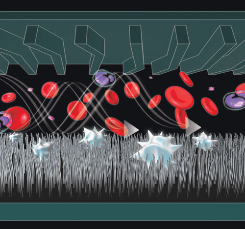
The NanoVelcro CTC technology is composed of two basic parts. First, a specialized nanostructure substrate is coated with “sticky” cell-capturing agents. This substrate consists of a silicon chip covered with nanopillars that interact with the microvilli of CTCs, attaching to them in a way similar to how the two sides of Velcro are joined together.
The second part of this system consists of a chaotic mixing chip, which is an overlaid microfluidic channel that creates a path that serves to increase CTC/substrate contact frequency.
“The device features high flow of blood samples, which travel at increased speed,” said Dr. Tseng. “The cells bounce up and down inside the channel, get slammed against the surface and get caught,” explained Clifton Shen, Ph.D., who collaborated with Dr. Tseng.
Dr. Tseng also presented data on the use of captured CTC cells.
“We could harvest the NanoVelcro-immoblized CTCs for subsequent molecular analyses (e.g., mutation sequencing). We found that the mutations identified in CTCs are responsible for patients’ disease progression.”
These results are part of three manuscripts currently in review. The new technology promises to significantly improve early detection of cancer metastasis and isolation of rare populations of cells, something not feasible with existing technologies, according to Dr. Tseng.
Regarding clinical applications, he plans to explore the use of the NanoVelcro CTC assay for monitoring therapeutic responses and resistance mechanisms in cancer patients who received targeted therapeutics.

NanoVelcro CTC technology contains a nanostructure substrate consisting of a silicon chip covered with nanopillars that interact with the microvilli of CTCs, attaching to them in a manner similar to how the two sides of Velcro are joined together. A chaotic mixing chip, which is an overlaid microfluidic channel that creates a path to increase CTC/substrate contact frequency, makes up the second part of the system. [UCLA]
Tubeless Microfluidic Systems
Albert Folch, Ph.D., associate professor in the bioengineering department of the University of Washington (Seattle), talked about an innovative tubeless microfluidic device, developed in his lab, for tissue-based chemotherapy drug-performance tests.
Research over the past two decades has provided new insights into the biology of tumors. They are no longer understood only as the sum of the characteristics of the cancer cells, but as complex tissues composed of different types of cells that interact with each other in many different ways.
Understanding the contributions of this tumor microenvironment to tumorigenesis is critical for the development of effective chemotherapy drugs, reported Dr. Folch.
Dr. Folch designed a microfluidic device aiming to mimic the actual environment of cancer cells when cultured outside the organism, while exposing them to a set of chemotherapy drugs to determine which one has the highest efficacy.
Dr. Folch initially faced a common challenge encountered by scientists designing microfluidic devices: how to connect these devices to the macroscopic world? Traditionally, macroscopic pumping systems with conventional connectors are used to push fluids in and out of the microfluidic devices, resulting in bulky systems.
“Most microfluidic devices are still designed with a lot of tubes coming out from them,” Dr. Folch said. “Three years ago, I told my students a lot of experimental failure was due to all these tubes because they generate bubbles and connection time becomes a very big issue.”
Thus, Dr. Folch’s microfluidic device differs from traditional ones. “It’s a modification of a 96-well plate. We cut out the center of 16 plates so that each of these acts as an inlet into the device, which is underneath the wells. The device is bonded underneath and each of these wells connects to one microchannel that leads to the center portion and that’s where we put our sample, our tissue,” he explained.
Advantages of such a tubeless device include a user-friendlier introduction of fluids, a diminished formation of bubbles, and less dead volume.
Dr. Folch’s long-term goal for his device is for “personalized chemotherapies.” So far, the tissues used have been animal biopsies that, once inside the device, are exposed to different drugs to see which work best.
Human-Microbial Molecular Interactions
Mahesh Desai, Ph.D., research associate at the center for systems biomedicine at the University of Luxembourg, presented his results with a microfluidics system called HuMiX, which he described as “a microfluidics-based in vitro co-culture device for investigating human-microbial molecular interactions.”
The main goal of Dr. Desai’s research is to understand how the different microbial communities (human microbiome) that inhabit our gastrointestinal tract interact and influence our health status.
“We have now seen that the microbes in our intestinal system have ten times more genes than our own body cells,” explained Dr. Desai.
Current work has identified differences in the human microbiome between healthy versus diseased individuals. However, it is still difficult to establish a causal link regarding what makes healthy people sick, he pointed out.
“The field is in desperate need of in vivo and in vitro methods that can study interactions of human bacteria and human body cells. We need to develop an individual system that can look at these interactions,” continued Dr. Desai.
“Therefore, we are designing a microfluidic system, HuMiX, to study human cell and microbial communities. This microfluidic system can combine models to test several hypotheses.”
HuMiX is a modular microfluidics-based device where human epithelial cell lines and human microbial communities can be grown in individual chambers, separated by a semi-permeable membrane. Their close proximity allows for molecular interactions between the two cell populations to occur and to be studied.
The device consists of three separate chambers (top, central, and bottom). The central chamber hosts the human epithelial cell lines, while the bottom chamber holds the microbial community. The upper chamber acts as a perfusion system and is separated from the middle chamber by a microporous membrane, which facilitates diffusion-based perfusion between chambers.
“The human contingents are perfused with standard human cell culture medium using a programmable syringe pump. The synthetic gut microbial communities are introduced in the bottom chamber after the epithelial cells undergo an enterocytic differentiation mimicking the intestinal epithelial cell barrier,” said Dr. Desai.
The device’s architecture allows researchers “to establish in vitro models for the human proximal colon, the entire human gastrointestinal tract, and human gastro-intestinal tissues,” allowing for the co-culture and study of human cells or tissues and their naturally co-existing mixed microbial communities. These cultures are further studied via microscopy and metabolomics approaches before an actual experiment is performed.
“Once both contingents reach a steady state, they are exposed to a particular experimental regime, and the devices are snap frozen and the respective cell contingents undergo omic analyses,” according to Dr. Desai.
In the near future, Dr. Desai aims to use HuMiX to address the role of microbial dysbiosis in the pathogenesis of different diseases.


